This is an automatically translated article.
Venous insufficiency of the lower extremities is a common disease in people who often have to stand for a long time in some special occupations. Color Doppler 4D ultrasound is an important subclinical measure for the diagnosis of venous insufficiency of the lower extremities to help detect the disease early.
1. Lower extremity venous insufficiency
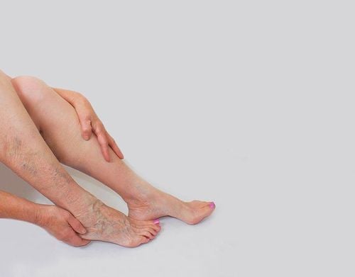
The lower extremity venous valve is a one-way valve that helps keep blood moving in a fixed direction toward the heart. Venous insufficiency of the lower extremities is a decline in the function of the venous system in the legs to return blood to the heart, leading to blood stagnation, causing distortions in surrounding tissue and changes in the surrounding tissues. on hemodynamics.
The cause of venous insufficiency of the lower extremities is unknown. However, risk factors for damage to the function of the one-way valve in the peripheral venous system include:
Working and working posture: Standing for too long or sedentary, sitting for a long time, carrying Heavy objects, ... are conditions that cause blood to pool in the legs and increase pressure in the veins in the lower extremities. The aging process due to age: Common in the elderly, especially with other diseases. Congenital valve defects People with multiple pregnancies, overweight and obesity, frequent constipation, tobacco use, stimulants, lack of exercise,...

Mang thai nhiều lần cũng làm tăng nguy cơ gây tổn thương tới chức năng của van một chiều
2. Color Doppler 4D . Ultrasound
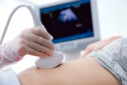
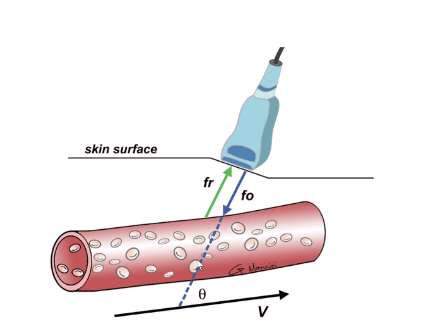
4D color Doppler ultrasound is an advanced ultrasound technique that uses continuous waves to detect flows, flow direction and velocity. Doppler ultrasound uses high-frequency waves that are not found in nature and has a high diagnostic value in evaluating vascular pathologies when vascular stenosis or occlusion is suspected, venous insufficiency or varicose veins are detected. ,...
The advantages of the Doppler ultrasound diagnostic method are:
Observe blood vessels and detect lesions accurately Ease of implementation Quick implementation time Non-invasive, no pain or discomfort for patients Ultrasonic waves are safe and do not affect health Cost saving compared to some other techniques.
3. Color Doppler ultrasound in the diagnosis of venous insufficiency of the lower extremities
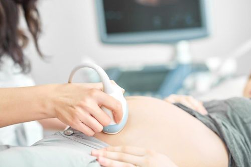
Doppler ultrasound is the preferred subclinical method in the diagnosis of venous insufficiency of the lower extremities. The person performing the ultrasound must be a doctor who is familiar with the anatomy of the superficial and deep veins. The ultrasound procedure for diagnosis of venous insufficiency of the lower extremities includes:
Investigation of the deep venous system: to find venous thrombosis and regurgitation. the great saphenous vein and the common femoral vein, and the junction of the minor and popliteal veins. The trunk of the saphenous veins: measure the diameter of the vein, find reflux in the trunk of the small saphenous vein and the vein of Giacomini, in the trunk of the great saphenous vein in the thigh, leg and anterior and posterior great saphenous branches.
Siêu âm doppler là biện pháp cận lâm sàng được ưu tiên sử dụng trong chẩn đoán suy van tĩnh mạch chi dưới
Superficial veins not belonging to the saphenous system: determine the location and flow of reflux. Direct and indirect perforating veins: determining the location and flow of reflux. Pulsed, color 4D doppler ultrasound is used to find spontaneous reflux or via squeezing maneuvers. In addition, it is necessary to use pressure test to find superficial vein thrombosis and use 2D ultrasound images to investigate.
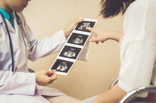
The diagnosis of venous insufficiency of the lower extremities should be based on the following criteria:
Detection of spontaneous regurgitation on color and pulsed Doppler ultrasonography or when performing squeezing or valsalava maneuvers. On color Doppler ultrasound, the signal is reversed from red to blue or vice versa. On pulsed Doppler ultrasound: regurgitation of superficial and deep veins >1000ms, femoral and popliteal venous regurgitation >500ms, transverse regurgitation >350ms In summary, limb venous insufficiency Below is common in people who often stand for a long time, sedentary and in some specific professions such as nursing, tailor, sales,... 4D Doppler ultrasound is the preferred diagnostic measure in valve failure. lower extremity veins. This is a subclinical method that has many advantages, is accurate, fast and does not affect the patient's health.
Vinmec International General Hospital with a system of modern facilities, medical equipment and a team of experts and doctors with many years of experience in medical examination and treatment, patients can rest assured to visit. examination and treatment at the Hospital.
Customers who need to be examined and treated at Vinmec International General Hospital, please register for an online examination on the Website for the best service.
Please dial HOTLINE for more information or register for an appointment HERE. Download MyVinmec app to make appointments faster and to manage your bookings easily.