This is an automatically translated article.
Birth defects in the fetus are always a nightmare for mothers, any fetus can have birth defects to different degrees, so the early detection of fetal malformations. is very important for proper handling and monitoring. Currently, ultrasound is one of the most effective, safest, and most widely used methods to detect fetal malformations, even at a very early age.1. What are fetal malformations?
Fetal malformations are abnormalities of the fetus that appear right in the fetus, which can be chromosomal abnormalities, morphology of one or more organs.In Vietnam, the rate of children with birth defects is about 3%, that is, 1 out of every 33 newborns is born with a birth defect. All pregnancies have a risk of fetal malformations, but the risk of fetal malformations is higher if the mother has factors such as:
Maternal age over 35 years old, the older the mother is at risk the higher the rate of fetal malformations The mother has a history of malformations of pregnancy, a history of multiple miscarriages, a family history of fetal malformations, the mother was infected with a virus during the first 3 months of pregnancy without being vaccinated (Rubella , Herpes, Cytomegalovirus ...), exposure to radiation, toxic chemicals. My mother has diabetes and smokes.
2. The role of ultrasound in diagnosing fetal malformations
Diagnosis of clinical fetal malformations is almost impossible, up to 90% of fetal malformations have no clinical manifestations. Fetal malformation ultrasound is a very safe, effective, easy to perform method with reasonable cost to diagnose and monitor pregnancy, as well as detect and monitor fetal malformations at other stages. At a very early age, fetal ultrasound under ideal conditions can accurately diagnose 85% to 90% of fetal malformations. The diagnostic value of fetal ultrasound depends greatly on the equipment and machinery, the level of analytical ability of the doctor doing the ultrasound, and the time of doing the fetal ultrasound.3. 3 landmark ultrasound fetal malformations pregnant women need to remember
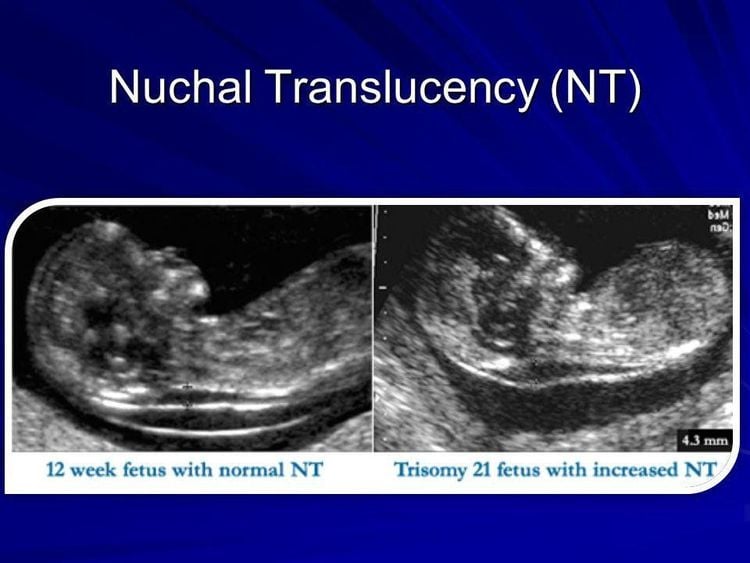
In the first 3 months of pregnancy, from 11 to 14 weeksIn the period from 12 weeks of age, the baby has developed relatively fully in terms of morphology and has reflexes such as bending and stretching. extremities... Therefore, this is one of the three important landmarks that experts recommend to perform. During this ultrasound, doctors will especially check and screen for early abnormalities of the brain, face, heart, digestive, urinary, extremities and the whole body as well as provide basic information of the patient. fetus, confirm the fetus is alive or not? See if the fetus is in the right position? How many pregnancy? Calculate the exact gestational age based on the length of the head of the buttocks.
Besides, pregnancy ultrasound within 12 weeks is a golden time to detect some fetal abnormalities if any such as: Down syndrome, Edward's syndrome... Because the baby in this stage is still still alive. The system is quite small, so a high-end 4D ultrasound system like the GE Voluson E10 will play a very important role in helping doctors at Vinmec detect more than 95% of anomalies during this period. Packed with the latest technologies, the GE Voluson E10 enables enhanced image quality and penetration for outstanding high-resolution images and easy operation.

Siêu âm thai 3 tháng đầu có vai trò quan trọng để chẩn đoán các bất thường nhiễm sắc thể của thai nhi
At this time, the fetus has basically fully developed its organs and organs, and the amount of amniotic fluid is also increased, allowing a good observation of the fetal morphology. This is the standard ultrasound time to evaluate the entire fetus.
This is an important milestone to detect most of the morphological abnormalities, confirm the abnormalities that were previously suspected, the final time for the decision to terminate the pregnancy if any (before the 28th week). Most morphological abnormalities can be diagnosed at this stage, the sonographer will look at the fetal parts in turn to evaluate the whole: Neurological abnormalities such as: Abnormalities neural tube, no brain, hydrocephalus, dilated ventricles, small brain, varicose veins of galen... Abnormalities of maxillofacial: Abnormalities are more clearly observed at ultrasound in the first month, especially Abnormalities in the eye can be observed. Cardiovascular abnormalities: At this stage, fetal ultrasound can clearly observe the heart and its structures, allowing the diagnosis of most abnormalities, including the most complex such as: atrioventricular, tetralogy of fallot, hypoplasia of the heart valves, Ebteins disease, right ventricular 2 outflow tract, cardiac arrhythmias... Thoracic abnormalities: diaphragmatic hernia, pulmonary cyst, pleural effusion, oliguria Abnormalities in the abdominal cavity, intestines and abdominal wall such as: esophageal stricture, gastric stenosis, hepatomegaly, splenomegaly, intestinal obstruction, umbilical hernia.... Abnormalities of the kidneys and urinary tract such as : Absence of kidney, polycystic kidney, urinary tract obstruction, abnormalities in bladder, urethra... Abnormalities in skeletal muscles and extremities: In addition to abnormalities detected in the first trimester ultrasound, stage This stage is more detailed observation of the fingers and feet, can easily detect defects such as: many fingers, crooked hands... Ultrasound in the last 3 months: Week 30 -32
This is the stage of pregnancy Children have fully matured in structure and developed rapidly. Fetal malformation ultrasound at this stage is mainly to evaluate fetal growth, fetal position, amniotic fluid, umbilical cord (and their abnormalities if any), the development of the uterus.. Fetal abnormalities that may be further detected or evaluated at this stage (compared to the mid-month period) include: Fetal malnutrition, genital abnormalities (position and movement) testicles, genital tumors, ovarian cysts, etc.), some abnormalities in the heart valves were more fully observed (heart tumor, stenosis of the heart valves, mitral aortic valve). , aortic abnormalities...), some brain abnormalities.

Lựa chọn địa chỉ chăm sóc thai sản và siêu âm thai để chẩn đoán dị tật thai nhi sớm và chính xác là hết sức quan trọng
Choosing a place for maternity care and pregnancy ultrasound to diagnose fetal malformations early and accurately is very important. It is very important to have a pregnancy ultrasound at the right time and periodically to detect fetal malformations early, so that appropriate monitoring and treatment measures can be taken (even a decision to terminate the pregnancy). The effectiveness of the ultrasound method for diagnosing fetal malformations depends a lot on the doctor's level, analytical ability, and modern equipment.
Vinmec International General Hospital is a high-quality medical facility that is highly appreciated by customers for its quality of examination and treatment as well as professional services. Vinmec has the most modern equipment system, with the most advanced ultrasound machines in the world and a team of experienced obstetricians in prenatal diagnosis and intervention to help monitor and detect early fetal malformations.
In addition, to protect the health of mother and baby comprehensively, Vinmec offers a variety of package Maternity services. With this package, the fetus will be tested Double Test or Triple Test to screen for fetal malformations to help detect dangerous fetal malformations that can be intervened early. Pregnant women will have regular prenatal check-ups with specialists, especially maternal thyroid screening tests in the first 3 months, Rubella tests. During pregnancy, if a pregnant woman encounters any abnormal health problems, she will be consulted and treated by a specialist doctor. After each visit, the doctors will analyze the ultrasound and test results and advise on health care, nutrition, and rest suitable for each pregnant woman, helping the mother and fetus to be healthy. well developed, uniform indicators.

Hình ảnh khách hàng được tư vấn sức khỏe sau khi khám thai tại Vinmec
Please dial HOTLINE for more information or register for an appointment HERE. Download MyVinmec app to make appointments faster and to manage your bookings easily.


