This is an automatically translated article.
The article is professionally consulted by Master, Doctor Tong Diu Huong - Radiologist - Department of Diagnostic Imaging - Vinmec Nha Trang International General Hospital.Ultrasonography of the eyeball, also known as ultrasound of the eye, is a useful method in diagnosing and determining the cause of eye diseases. The doctor will appoint the patient to perform an eye ultrasound when he has just injured the eye or has eye diseases for which the cause is unknown.
1. What is ocular ultrasound?
Ultrasonography of the eyeball is a method commonly used today, valuable in helping doctors detect diseases in the patient's eyes and the causes of that damage in the most accurate way. . The ocular ultrasound method uses high-frequency sound waves to create detailed images of the patient's eye and orbital structure, so that the doctor will have a more detailed view of the internal structure. in the eye and accurately assess the disease state.2. When is an ultrasound of the eyeball needed?
In case the patient has eye problems of unknown cause or has an eye injury accident, the doctor will prescribe an ultrasound of the eyes and eye sockets. In addition, eye ultrasound can also help detect some conditions such as:
Retinal detachment
Tumor or cell proliferation in the eye
● Foreign object in the eye
● Glaucoma
● Cataracts
● Lens implants
● Measure the thickness and spread of the cancerous tumor.
Một số bệnh lý về mắt được chỉ định siêu âm nhãn cầu
3. Attention when conducting eyeball ultrasound
Unlike some other ultrasounds that need to be prepared before the ultrasound, the ophthalmic ultrasound is a very simple method and the patient does not need to prepare anything, the ultrasound process will feel no pain. To help the ultrasound process go smoothly, the patient should use eye drops to help the eyes relax and clean more.
After the end of the eyeball ultrasound, the patient's eyes will be temporarily limited in vision for a short time due to the influence of the ultrasound process, so it is advisable to have a family member accompany and return to make sure. safe.
In addition, to protect the cornea from damage, after the ocular ultrasound, the patient should not rub the eye until the anesthetic wears off.
4. Eyeball ultrasound procedure
A-scan and B-scan are the whole eyeball ultrasound process that will take place within 20-30 minutes. The doctor will conduct an eye measurement (A-scan) and then a B-scan to clearly see the space inside the eye.
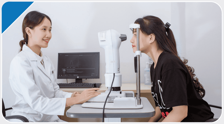
● A-scan : Playing an important role, helping to measure the size of the eyes, to perform this procedure, the patient will sit upright in a chair, put his chin on a specialized measuring device and look straight ahead, A lubricant probe will be placed on the front part of the eye during the scan.
● B-scan: In case the patient has some eye disease that makes it difficult for the doctor to see the back of the eye or has cataracts, it is necessary to conduct a B-scan immediately. A B-scan will help see the space behind the eye and diagnose tumors, retinal detachments, and other conditions. The patient will be asked to close their eyes so that the doctor applies the gel to the eyelids and moves the eyeballs in different directions according to the doctor's instructions.

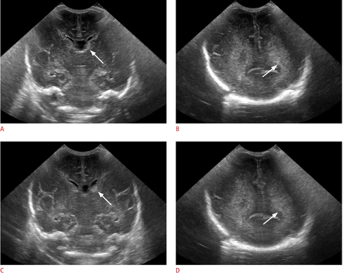
Hình ảnh siêu âm nhãn cầu
5. What does an ultrasound of the eyeball show?
Ultrasound image of the eyeball will help the doctor to accurately diagnose the situation and give the final result. The doctor will review the patient's eye measurements to make sure the eye measurements are within the normal range and then look at the B-scan results for information about the eye's structure. If the results are abnormal, your doctor will look for the specific cause.
Some eye conditions can be detected by B-scan including:
Retinal detachment
Damaged tissue or trauma to the orbit
● Cancer of the retina, located below the retina or other parts of the eye
Foreign body in the eye
● Swelling
Vitreous hemorrhage (blood flowing into the vitreous cavity at the back of the eye)
An ultrasound of the eyeball is a simple, performed procedure The operation is quick and gentle, so the patient will not feel any pain or experience any complications after an ultrasound of the eyeball. In order to keep the eyes healthy and bright, as soon as they see any abnormality in the eyes, the patient should go to the hospital to be examined by a specialist doctor, accurately diagnose the condition and have a timely treatment plan.
Eye Department of Vinmec International General Hospital and a team of leading ophthalmologists provide eye examination, treatment and surgery services with high quality, intensive ophthalmological techniques that bring efficiency and safety. care for customers.
A team of experts, leading ophthalmologists with rich experience in many fields
The first and only hospital in Vietnam committed to the quality of cataract surgery through providing boxes and cards ensuring information and quality of artificial lenses, ensuring European standards for using surgical supplies
One of the first private hospitals in the North to deploy routine corneal transplantation In the treatment of diseases
● Vinmec uses Ortho-K contact lens products from the United States and Japan with world leading quality and reputation.
To register for examination and treatment of ophthalmic diseases at Vinmec International General Hospital, you can contact Vinmec Health System nationwide, or register online HERE.

Khám mắt tại Bệnh viện Vinmec
SEE MORE
Eye surgeries performed at Vinmec hospital How is conjunctivitis transmitted? What to avoid when sick? ORTHO K - Solution to treat Myopia, Astigmatism without surgery


