This is an automatically translated article.
The article is professionally consulted by Master, Doctor Nguyen Quang Duc - Doctor of Nuclear Medicine - Department of Diagnostic Imaging and Nuclear Medicine - Vinmec Times City International General Hospital.To examine the abdominal aortic aneurysm, the doctor usually assigns the patient to perform a CT scan of the aorta - iliac with iodine contrast injection. This is an easy-to-implement and highly accurate imaging method.
1. An overview of abdominal aortic aneurysm
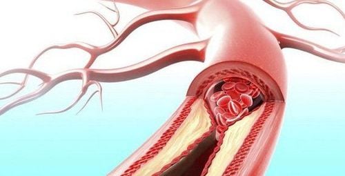
The aorta is the body's largest artery, originating from the heart, running in an arc in the chest, through the diaphragm down to the abdomen. The aorta divides into branches, which supply blood to the organs of the body. There are many pathologies that occur in the aorta, of which aortic aneurysm is a common condition. Aortic aneurysms include thoracic aortic aneurysms and abdominal aortic aneurysms.Aortic aneurysms progress when the aortic wall is weakened by risk factors such as: smoking, aging (more common in people over 60 years old), male gender, high blood pressure, atherosclerosis arteries, family history, certain genetic diseases, trauma to the abdomen or chest,...
Symptoms of abdominal aortic aneurysm are often atypical, mainly abdominal pain, back pain, and mass in the abdomen pulsating in accordance with the heart rate, occlusion of the lower extremities. When the abdominal aorta ruptures, the symptoms will be overwhelming including abdominal pain, abdominal distention, bleeding in the abdomen,... If not timely intervention, the patient will definitely die.
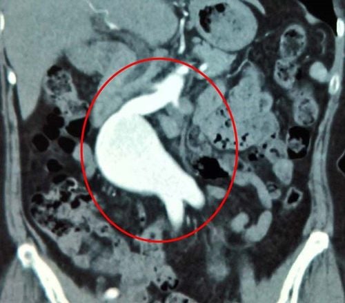
Abdominal aortic aneurysms can be diagnosed through a CT scan of the aorta-pelvis with iodine contrast injection.
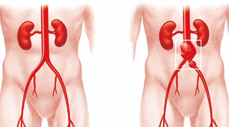
Động mạch chủ là động mạch lớn nhất của cơ thể
2. Procedure for CT scan of the aorta - pelvis with iodine contrast injection
CT scan of the iliac aorta with iodinated contrast is a minimally invasive arteriole imaging and examination technique.When applying this method, the doctor will put iodine contrast medicine into the patient's body through an intravenous line. Contrast drugs temporarily change the way X-rays interact, make structures visible, improve visualization of organs, blood vessels or tissues, help doctors diagnose your health condition patient correctly.
2.1 Indications/contraindications
This method is indicated for the examination of abdominal aortic aneurysms:● Helps determine the diameter of the aneurysm; bulge shape (vesicle or rhombus);
● Location of abdominal aortic aneurysm; assessment of periarterial fat;
Additional diagnostics for angiography and Doppler ultrasound;
● Consider pre-treatment of aneurysms (by surgery or endovascular intervention) and emergency visit for abdominal aortic aneurysms.
Contraindication to CT scan of the aorta - pelvis with iodine contrast injection: It should be considered in cases where the patient is pregnant, has renal failure or is allergic to contrast agents,...
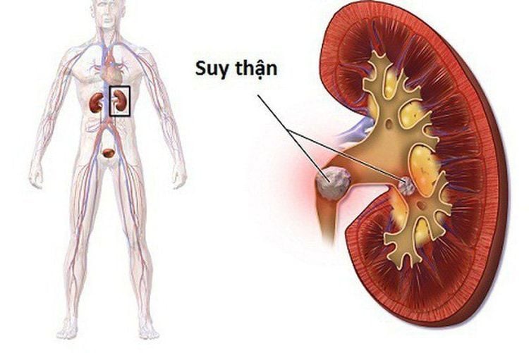
Bệnh nhân suy thận cần xem xét kĩ càng khi chụp CLVT động mạch chủ - chậu có tiêm thuốc đối quang i ốt
2.2 Preparation for implementation
Implementation personnel: Specialist doctors, nurses and radiology technicians; ● Technical facilities: Computerized tomography machine with 8 rows or more, specialized electric pump, film, film printer, image storage system;● Medical supplies: 10 and 20ml syringes, 18 - 20G needles, syringes for electric pumps, skin-mucous antiseptic solutions, water-soluble iodine contrast agents, physiological saline or distilled water, cotton, surgical gauze, gloves, hats, surgical masks, medicine boxes, first-aid equipment for contrast agents;
● Patient: The procedure is explained in detail (purpose, procedure, risk of complications); remove earrings, necklaces, hairpins, hearing aids, dental instruments if any; fast, drink 4 hours before the scan (can drink less than 50ml of water); for patients who are overstimulated, do not lie still,... use sedatives;
● Test card: There is a slip to order a computerized tomography scan.

Trường hợp bệnh nhân bị kích thích quá mức, bác sĩ có thể sử dụng thuốc an thần
2.3 Implementation process
● Examining the patient: Reviewing the medical record, looking for signs of contraindications to intravenous iodine contrast; prepare an intravenous line with an 18G needle and refer to previous imaging findings if available;● Patient position: Lying supine on the imaging table, hands on the head and 120ml of iodine contrast agent injected at the rate of 3ml/s using an electric syringe;
Capture positioning;
● Before injection: Do not inject iodinated contrast agent - take according to the required technical procedure;
● Arterial phase: 20 seconds after the start of iodine contrast injection - take according to the required technical procedure;
● Reconstruction according to technique, using MPR, MIP, VR, 3D software to reconstruct images of aorta, aneurysm, related positions with iliac and renal arteries.
After completing the imaging, the body will absorb or excrete the iodinated contrast agent through the urinary or gastrointestinal tract.
2.4 Evaluation of results
● Computed tomography images show the anatomical structures of the aorta - iliac system;● Review images on 2D cross-sections, supplemented with 3D reconstructions.

Hình ảnh chụp cắt lớp vi tính hiển thị được các cấu trúc giải phẫu của hệ thống động mạch chủ - chậu
2.5 Complications and how to deal with them
● Error to repeat the technique: The patient did not keep motionless during the computed tomography scan, the resulting image was not clear;● Digestive disorders or diarrhea : Caused by drinking a lot of water. With this complication, only medical treatment is needed;
● Complications related to iodinated contrast agents: Mild reactions (nausea and vomiting, itching, headache, hot flushes, skin rash or mild urticaria), moderate reactions (wheezing, difficulty breathing, severe skin rash or hives, irregular heartbeat, low or increased blood pressure ) and severe reactions (difficulty breathing, convulsions, cardiac arrest, swelling of the throat or other body parts, low blood pressure serious). Depending on the individual case, the doctor will have an appropriate treatment plan.
Patients assigned to perform a CT scan of the aorta - pelvis with iodine contrast injection should strictly follow all instructions of the doctor to make an accurate, quick diagnosis and limit the risk of complications.
German doctor has more than 12 years of experience in the field of Diagnostic Imaging. Recently, the Doctor has been assigned to specialize in the field of Nuclear Medicine. Currently, the doctor is studying for an intensive course in Nuclear Medicine at Stanford Hospital, USA
All questions need to be answered by a specialist doctor as well as customers wishing to examine and treat at the hospital. You can contact Vinmec Health System nationwide or register online HERE.
SEE MORE
Stent graft for aneurysm and aortic dissection at Vinmec Times City Surgery to replace the abdominal aorta below the kidney How does the aortic aneurysm affect other organs?


