
12 prenatal diagnostic tests (PND) pregnant women need to know
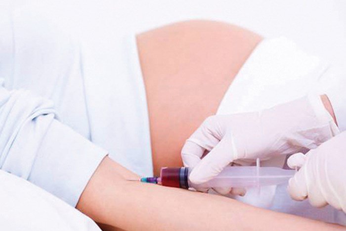
In order for your baby to be born healthy and have a normal life like everyone else, a pregnant mother needs to do a lot of things. In addition to pre-vaccination, during pregnancy to help monitor the periodic development of the fetus, pregnant women should perform prenatal screening to determine the risk of birth defects. Let's find out what are 12 prenatal diagnostic tests through the following article.
1. Karyotype Test – Chromosome Set Up The karyotype test looks at your size, shape and number of chromosomes. Chromosomes are the parts of your cell that contain your genes. Genes are part of DNA passed down from your mother and father. They carry information that defines your unique features, such as height and eye color.
The normal person has 46 chromosomes, divided into 23 pairs, in each cell. One of each pair of chromosomes comes from your mother, and the other from your father.
If you have more or less than 46 chromosomes, or if there are any abnormalities in the size or shape of the chromosomes, it could mean that you have an inherited disease. Karyotype testing is often used to help find genetic defects in a developing baby.
2. QF-PCR Assay Quantitative Fluorescence PCR (QF-PCR) is an alternative method in which markers of DNA polymorphisms on chromosomes, are used to determine the presence of different alleles. .
Test based on the use of polymorphic small tandem repeat (STR) markers with information and parental DNA availability, used for prenatal and postnatal diagnosis of heterozygotes multiples of chromosomes 13, 18, 21, X and Y. DNA was isolated from fetal cells from amniotic fluid samples, chorionic villus samples, and fetal trophoblast cells from endometrial lavage fluid. Bowel and neonatal blood were both used to investigate chromosomal copy number variations.
The QF-PCR assay using fluorescently labeled STR primers was analyzed after fragment length separation in capillary gel electrophoresis. Determination of the biological origin of the aneuploidy or of a post-mitotic origin can also be done in most cases. Although prenatal testing is complicated by limited sample sizes, variable sample quality, mosaicism, and maternal cell contamination - the use of parental samples and other measures can overcome most of these limitations.
The QF-PCR technique serves as a very useful preliminary test to alleviate parental anxiety in the short term and to accelerate therapeutic intervention.

3. FISH Test Fluorescence in situ hybridization (FISH) is a type of cytogenetic technique that uses fluorescent probes that link sections of chromosomes to show high levels of sequence complement.
Fluorescence microscopy can be used to find where the fluorescent probe is attached to the chromosome. This technique offers a new way for researchers to visualize and map the genetic material in an individual cell, including specific genes or sections of genes. It is an important tool for understanding many types of chromosomal abnormalities and other genetic mutations. Unlike most other techniques used to study chromosomes, FISH does not need to be performed on actively dividing cells, which makes it a very versatile procedure.
4. Thalassemia Gene Test Thalassemia is a group of inherited blood disorders that can be passed on from parents to their children and affect the amount and type of hemoglobin the body produces.
Hemoglobin (Hb or Hgb) is a substance found in all red blood cells (RBCs). It is important for the proper function of red blood cells because it carries the oxygen that red blood cells provide throughout the body. A part of hemoglobin called heme is the molecule that has iron in it...
Alpha thalassemia is sometimes confused with iron deficiency anemia because both disorders have red blood cells that are smaller than normal (microcytic). ). If someone has thalassemia, his or her iron levels are not expected to be low. Iron therapy will not help people with alpha thalassemia and can lead to iron overload, which can damage organs over time.
Evaluation of hemoglobin (Hb) disease (hemoglobin electrophoresis). This test evaluates the type and relative amount of hemoglobin present in red blood cells. Hemoglobin A (Hb A), which includes both alpha and beta globin, is the type of hemoglobin that normally makes up 95% to 98% of hemoglobin in adults. Hemoglobin A2 (HbA2) typically makes up 2% to 3% of hemoglobin in adults, while hemoglobin F usually makes up less than 2%.
Beta thalassemia upsets the balance of beta chain formation and alpha hemoglobin and causes an increase in those small hemoglobin components. Therefore, people with beta thalassemia major often have an Hb F ratio. People with beta thalassemia major usually have a higher Hb A2 ratio. Hb H is a less common form of hemoglobin that can be seen in some cases of alpha thalassemia. Hb S is the hemoglobin that is more common in people with sickle cell disease.
Evaluation for hemoglobin (Hb) disease is used for state-regulated neonatal hemoglobin screening and prenatal screening when parents are at high risk for hemoglobin abnormalities.
Amniotic fluid genetic testing is used in rare cases where the fetus is at higher risk of thalassemia. This is especially important if both parents are likely to carry the mutation because it increases the risk that their child may inherit a combination of abnormal genes, which causes a more severe form of thalassemia.

5. Duchenne Muscular Dystrophy Gene Testing Genetic testing involves analyzing the DNA of any cell (usually blood cells are used) to see if there is a mutation in the dystrophin gene, and if so, the exact location where it happened.
Usually, genetic diagnosis is indicated for patients with high serum CK levels and clinical findings of nasal dystrophy. The diagnosis is confirmed if a mutation of the DMD gene is identified. Genetic analysis is aimed primarily at finding large deletions/duplications (70% to 80% of cases have these mutations). If the initial gene analysis was negative, then analysis for micro and small deletion/duplicate mutations was followed.
Female relatives of men and men with DMD can undergo DNA testing to see if they are carriers. DMD carriers can pass the disease on to their sons and carrier status to their daughters. In rare cases, girls and women with DMD can develop symptoms of DMD on their own, such as muscle weakness and heart problems. These symptoms may not appear until adulthood.
Several experimental drugs are currently being developed to treat DMD that require knowledge of a person's exact genetic mutation, so genetic testing has become important not only for diagnosis but also possible for future treatments.
6. Hemophilia gene test – Hemophilia 6.1. Complete blood count (CBC) This common test measures the amount of hemoglobin (the red pigment inside red blood cells that transport oxygen), the size and number of red blood cells, and the number of different types of white blood cells and platelets. Various are found in the blood. CBC is normal in people with hemophilia. However, if a person with hemophilia bleeds abnormally heavily or bleeds for a long time, the hemoglobin and red blood cell count may be low.
6.2. Partially Activated Thromboplastin Time Test (APTT) This test measures the time it takes for the blood to clot. It measures the clotting ability of factors VIII (8), IX (9), XI (11) and XII (12). If any of the clotting factors are too low, the blood will take longer than usual. The results of this test will show a longer clotting time in people with hemophilia A or B.
6.3. Prothrombin time (PT) test This test also measures the time it takes for the blood to clot. It mainly measures the clotting ability of factors I (1), II (2), V (5), VII (7) and X (10). If any of these factors are too low, the blood will take longer than usual. The results of this test will be normal for most people with hemophilia A and B.
6.4. Fibrinogen test This test also helps doctors assess a patient's likelihood of blood clots forming. This test is ordered along with other coagulation tests or when the patient has an abnormal external PT symbol or an internal APTT symbol result, or both. Fibrinogen is another name for clotting factor I (1).
6.5. Coagulation factor testing A clotting factor test, also known as a factor test, is required to diagnose a bleeding disorder. This blood test shows the type of hemophilia and its severity. It is important to know the type and severity to plan the best treatment.

7. Down Syndrome Testing Down syndrome is a disorder that causes intellectual disability, distinctive physical features, and various health problems. These can include heart defects, hearing loss, and thyroid disease. Down syndrome is a chromosomal disorder.
Chromosomes are the parts of your cell that contain your genes. Genes are part of DNA passed down from your mother and father. They carry information that defines your unique features, such as height and eye color.
The normal person has 46 chromosomes, divided into 23 pairs, in each cell. One of each pair of chromosomes comes from your mother, and the other from your father. In Down syndrome, there is an extra copy of chromosome number 21.
Extra chromosomes change the way the body and brain develop. Down syndrome, also known as trisomy 21, is the most common chromosomal disorder in the United States.
In two rare forms of Down syndrome, called mosaic trisomy 21 and translocation trisomy 21, the extra chromosome is not present in all cells. People with these disorders often have few of the features and health problems associated with the common form of Down syndrome.
Down syndrome screening tests show whether your baby is more likely to have Down syndrome. Other types of tests confirm or rule out the diagnosis.
8. Testing for Edwards Syndrome 1 in every 5,000 children is diagnosed with Trisomy 18, also known as Edwards syndrome. Normally, a person has 23 pairs of chromosomes. Chromosomes are packets of genetic information, made up of DNA, that contain the instructions the body uses to build a person. Chromosomes have 23 pairs, with most people having a total of 46 chromosomes. Trisomy 18 is caused when a person has three copies of chromosome 18 instead of the usual two, for a total of 47 chromosomes.
This extra chromosome affects the baby's development, leading to a number of medical problems that can include: heart defects, gastrointestinal abnormalities, cleft lip, contractures (abnormal bending) , vision and hearing problems, developmental delays before and after birth, seizures, and hypotonia (weak muscles). All infants who survive Trisomy 18 have significant (often severe) intellectual disability.
9. Turner Syndrome Testing Prenatal screening tests that evaluate the baby's DNA in the mother's blood (prenatal cell-free DNA screening or noninvasive prenatal screening) may also give found an increased risk of Turner syndrome. However, karyotype should be done during pregnancy or after delivery to confirm the diagnosis.
A pregnancy and childbirth specialist (obstetrician) may ask if you are interested in additional tests for prenatal diagnosis of your baby. One of two procedures may be performed to check for Turner syndrome preoperatively:
Chorionic villus sampling. This involves taking a small piece of tissue from the developing placenta. The placenta contains the same genetic material as the baby. Chorionic villus cells may be sent to a genetics lab for chromosome studies.
Amniocentesis. In this test, a sample of amniotic fluid is taken from the uterus. The baby sheds the cells into the amniotic fluid. The fluid may be sent to a genetics lab to study the baby's chromosomes in these cells.
10. Genetic testing for DiGeorge Syndrome The diagnosis of DiGeorge syndrome (deletion 22q11.2) is primarily based on a laboratory test that can detect a deletion on chromosome 22.
Doctor will probably order this test if your child has: A combination of problems or medical conditions that suggest deletion syndrome 22q11.2 Heart defects, as some heart defects are commonly associated with deletion syndrome 22q11.2 In some cases, a child may have a combination of conditions suggestive of 22q11.2 deletion syndrome, but laboratory testing does not indicate a chromosomal deletion. 22. Although these cases present a diagnostic challenge, the coordination of care to address all medical, developmental, or behavioral problems is likely to be similar.
11. Cytomegalovirus DNA testing Cytomegalovirus (CMV) is a common virus that usually causes no symptoms or only mild illness. The CMV test detects antibodies in the blood that the body produces in response to an infection or directly detects CMV.
The Cytomegalovirus (CMV) test is not used to check everyone for CMV infection. It can be used to help diagnose active, reactivated, or past CMV infection under certain circumstances, such as:
Some pregnant women or those in decline immunity to signs and symptoms People who may receive an organ or bone marrow transplant Newborns with certain congenital (congenital) abnormalities Several different testing methods may be used depending on for testing purposes: Antibody (serology) test
12. Uniparental Disomy 15 (UPD) test - Angelman syndrome and Prader Willi syndrome Prader Willi syndrome for short PWS or also known as Disomy 15 (UPD 15), a very rare syndrome, is caused by the loss of function of a gene on the long arm of chromosome 15. The frequency is from 1 in 10,000 to 1 in 30,000 newborns.
A possible cytogenetic technique is tape-stretch chromosomal analysis to detect deletions of 15q11 - q13 and other associated chromosomal abnormalities.
FISH technique is a hybrid technique between cell and molecular genetics commonly used to detect loss of chromosome 15 region of band q11 - q13 of paternal origin.
BoBs (Bacs-on-Beads) and aCGH techniques are used to detect small deletions that include sites on chromosome 15.
NIPT (mother's blood prenatal screening) can screen fetus with Prader Willi syndrome.

Vinmec currently has many maternity packages (12-27-36 weeks), in which the 12-week maternity package helps monitor the health of mother and baby right from the beginning of pregnancy, early detection and timely intervention. health problems. In addition to the usual services, the maternity monitoring program from 12 weeks has special services that other maternity packages do not have such as: Double Test or Triple Test to screen for fetal malformations; Quantitative angiogenesis factor test to diagnose preeclampsia; thyroid screening test; Rubella test; Testing for parasites transmitted from mother to child seriously affects the baby's brain and physical development after birth.
For more information about the 12-week maternity package and registration, you can contact the clinics and hospitals of Vinmec health system nationwide.
Để đặt lịch khám tại viện, Quý khách vui lòng bấm số HOTLINE hoặc đặt lịch trực tiếp TẠI ĐÂY. Tải và đặt lịch khám tự động trên ứng dụng MyVinmec để quản lý, theo dõi lịch và đặt hẹn mọi lúc mọi nơi ngay trên ứng dụng.
Dịch vụ từ Vinmec
-
 Các xét nghiệm đặc biệt để theo dõi sức khỏe thai nhi
Trong thời gian mang thai, để đảm bảo cho người mẹ và thai nhi khỏe mạnh, thai phụ cần thực hiện các hình thức xét nghiệm khác nhau. Dưới đây,là các thông tin hữu ích về những xét nghiệm đặc ...
Đọc thêm -
 Những điều cần biết về sinh thiết gai nhau
Sinh thiết gai nhau (CVS - Chorionic Villus Sampling) là thao tác kỹ thuật áp dụng trong sản khoa: lấy một mẫu tế bào là phần màng đệm bao bọc quanh phôi thai, còn gọi là gai nhau để tiến ...
Đọc thêm -
 Vì sao nên sàng lọc kháng thể trước khi sinh?
Khi bạn chuẩn bị làm mẹ, một trong những xét nghiệm mà bạn sẽ phải làm đó là sàng lọc kháng thể trước sinh. Xét nghiệm trước sinh này nhằm tìm kiếm những kháng thể trong cơ thể bạn, một ...
Đọc thêm -
 Thai 13 tuần 2 ngày độ dày da gáy 0,2cm có cần xét nghiệm lại không?
Chào bác sĩ! Em 34 tuổi mang thai con thứ 3, em có thai được 13 tuần 2 ngày. Em đi siêu âm bác sĩ nói độ dày da gáy của thai nhi là 0,2cm là cao, mà trước đó ...
Đọc thêm -
 Có thể chữa khỏi bệnh bạch cầu tủy mạn hoàn toàn không?
Chào bác sĩ, Em bị chẩn đoán bệnh bạch cầu tủy mạn. Bác sĩ cho em hỏi có thể chữa khỏi bệnh bạch cầu tủy mạn hoàn toàn không? Em cảm ơn.
Đọc thêm