
The article is professionally consulted by Specialist Level I, MD. Le Hong Lien, Department of Obstetrics and Gynecology, Vinmec Central Park International General Hospital.
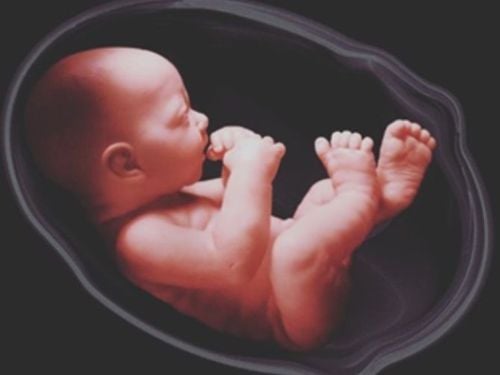
Amniotic fluid serves to nourish and protect the fetus throughout pregnancy. By examining the color and volume of the amniotic fluid, doctors can assess the health status and certain conditions of the fetus.
1. What color is amniotic fluid?
In the early stages of pregnancy, amniotic fluid is clear and colorless. As the fetus grows, the color of the amniotic fluid gradually becomes cloudy white due to the presence of vernix caseosa (a substance that coats the baby's skin to protect it, similar to fat). When the fetus reaches full maturity (from the 38th week), the amniotic fluid will have a cloudy white color, resembling rice water.
Abnormal colors of amniotic fluid can be a warning sign of potential health issues concerning the fetus.
- Amniotic fluid that is yellow-green: may indicate fetal hemolysis or intrauterine growth restriction.
- Cloudy or greenish amniotic fluid mixed with meconium suggests severe fetal distress in the womb, posing a threat to the baby's life.
- If the amniotic fluid appears cloudy with a purulent consistency and has a foul odor, this indicates an infection of the amniotic fluid, with a high risk of the baby contracting an infection in utero, etc…
- Brownish-red amniotic fluid may suggest that the fetus has experienced intrauterine fetal demise.
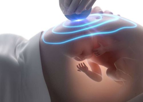
The color of the amniotic fluid can be observed through amnioscopy in cases where the cervix is dilated more than 1 cm or by performing amniocentesis through the abdominal wall. When the membranes are ruptured artificially or naturally, the color of the amniotic fluid can be accurately and clearly assessed.
2. Abnormalities in amniotic fluid volume
The volume of amniotic fluid gradually increases from 250 to 800 ml between 16 and 32 weeks, stabilizing at this level until 36 weeks, and then gradually decreasing to about 500 ml by the time of delivery. In practice, ultrasound is used to assess whether the amniotic fluid is low or high; there are two methods of measurement: either by directly measuring a specific area of amniotic fluid (measuring the largest diameter of the cavity or the volume of the cavity) or by dividing the uterine cavity into four sections and evaluating the amniotic fluid in each section, then summing the results to obtain the amniotic fluid index (AFI). Clinical assessment is based on palpating the abdomen, where the fundal height may not correspond to the gestational age (lower when there is oligohydramnios or higher when there is polyhydramnios), and whether the abdomen feels firm or lax. However, these signs can also be due to other factors, such as a large fetus, fetal malnutrition, multiple pregnancies, or the mother's body weight being too low or too high. Therefore, ultrasound remains the most accurate method of assessment.
2.1. Polyhydramnios
Polyhydramnios is the condition characterized by an excess accumulation of amniotic fluid. A pregnant woman is diagnosed with polyhydramnios when the amniotic fluid index (AFI) measured by ultrasound exceeds 25 cm, and the largest amniotic cavity is greater than 8 cm. Polyhydramnios is often observed in multiple pregnancies and certain abnormalities of the fetal central nervous system, such as hydrocephalus, anencephaly, meningocele, and spina bifida, etc,... It can also be caused by pathological conditions of the amniotic membranes, placenta, umbilical cord, placental edema, fetal macrosomia, maternal conditions such as diabetes, or it may be idiopathic.
In practice, about two-thirds of polyhydramnios cases have no identifiable cause. Potential causes of polyhydramnios in mothers may include:
- Maternal diabetes: Polyhydramnios is found in 10% of pregnant women with diabetes, especially in the third trimester. When a mother has diabetes and her blood sugar levels are not well controlled, the baby may produce more urine, leading to increased amniotic fluid. Therefore, controlling blood sugar levels can help reduce the amount of amniotic fluid.
- Maternal hypertonia: However, this condition is quite rare during pregnancy.
- Multiple pregnancies: polyhydramnios can occur due to an imbalance in the metabolic exchange between two fetuses (one fetus may have less amniotic fluid while the other has more).
- Fetal abnormalities: The fetus may stop the process of swallowing amniotic fluid and urinating, leading to an excess of amniotic fluid. This condition can occur with fetal malformations such as cleft lip and palate or pyloric stenosis, etc,...
- Other factors that may contribute to increased polyhydramnios include fetal anemia, intrauterine infections, and blood group incompatibility between the mother and the baby.
Effects of polyhydramnios on fetal development and health: polyhydramnios occurs in about 1% of pregnancies, and if it is due to congenital anomalies or malformations, the prognosis is very poor. Polyhydramnios allows the fetus to move more freely in the uterus, which can lead to complications such as umbilical cord entanglement, cord knots, and abnormal fetal positions. It causes the mother's abdomen to become distended, making it difficult for her to breathe, and increases the risk of uterine contractions and preterm labor, which raises the risk of neonatal mortality. During labor, polyhydramnios can lead to prolonged labor, increasing the risk of fetal distress and uterine atony, which can result in postpartum hemorrhage. Additionally, polyhydramnios may cause sudden rupture of membranes, with complications such as placental abruption, cord prolapse, abnormal fetal positions, and amniotic fluid embolism. These conditions pose a threat to the lives of both the mother and the baby. Of course, these risks can vary depending on the severity and underlying cause of the polyhydramnios. Immediately after the baby is born, the excess fluid is released, and the mother often feels much more comfortable.
In most cases of mild polyhydramnios, there is no cause for concern. The doctor will advise regular check-ups and may prescribe diuretics. If an infectious cause can be identified, it can often be treated safely with antibiotics for the fetus.
For severe polyhydramnios, if there are signs indicating potential issues with the fetus, close monitoring is necessary. The doctor will carefully monitor your amniotic fluid levels, and if they increase too rapidly, you may need surgery or amniocentesis to reduce the fluid.
Additionally, the mother should have regular check-ups to monitor her condition and may need to be hospitalized and treated immediately if severe symptoms arise, such as difficulty breathing, chest tightness, rapid and noticeable abdominal enlargement, or sudden pain.
2.2. Oligohydramnios
Oligohydramnios is the condition characterized by a lower than normal amount of amniotic fluid, defined as an amniotic fluid index (AFI) of less than 5 cm with intact membranes. It is often associated with abnormalities of the fetal urinary and digestive systems. Oligohydramnios can also occur in cases of maternal malnutrition, fetal growth restriction, post-term pregnancy, premature rupture of membranes, and early rupture of membranes, etc,...
Effects of oligohydramnios on fetal development: The impact of low amniotic fluid levels on fetal development depends on the condition, underlying cause, and gestational age.
- If a pregnant woman experiences oligohydramnios in the first trimester or a few weeks thereafter, there is a risk of miscarriage or stillbirth. Additionally, this condition can affect the development and function of the fetus's lungs.
- If oligohydramnios occurs in the third trimester, the mother may feel somewhat relieved, as it typically does not lead to severe complications. The health of both the mother and the fetus will be closely monitored, and intravenous fluids may be administered to supplement amniotic fluid. However, oligohydramnios at this stage can lead to abnormal fetal positioning, as there may not be enough amniotic fluid to allow the fetus to turn head-down.
- Oligohydramnios can also cause premature rupture of membranes. If the membranes rupture and lead to oligohydramnios before labor begins or when contractions are just starting, it can result in amniotic infection, which may lead to fetal infection and uterine infection, etc,... posing risks to both the mother and the baby. The doctor will assess the risk of amniotic infection and the condition of the fetus to determine whether early intervention is necessary.
To treat oligohydramnios, the doctor will take a medical history and perform a vaginal fluid test (Nitrazine test) to rule out amniotic fluid leakage or rupture. Following this, a prenatal ultrasound will be conducted to investigate and identify any morphological abnormalities in the fetus, particularly concerning the fetal urinary system, such as renal dysplasia or urinary tract obstruction. The patient will be informed about the benefits and potential complications of the procedure, and an amnioinfusion may be performed if the low amniotic fluid levels hinder the assessment of fetal morphology. Additionally, amniotic fluid may be collected for immunological and genetic testing to relieve pressure on the umbilical cord and allow for fetal movement. In cases where there is also intrauterine growth restriction, fetal heart monitoring, Doppler ultrasound (AFI, middle cerebral artery Doppler), and obstetric monitoring, etc,... may be recommended.

Abnormalities in the volume and color of amniotic fluid can lead to serious consequences for fetal health and can also cause anxiety for pregnant women. Therefore, in addition to maintaining a proper diet and staying hydrated, expectant mothers should not forget to attend regular prenatal check-ups as advised by their obstetrician, in order to detect and treat any potential abnormalities that may arise during pregnancy in a timely manner.
Please dial HOTLINE for more information or register for an appointment HERE. Download MyVinmec app to make appointments faster and to manage your bookings easily.