This is an automatically translated article.
The article was professionally consulted by Specialist Doctor I Le Hong Lien - Department of Obstetrics and Gynecology - Vinmec Central Park International General Hospital. Doctor Lien has over 10 years of experience as a radiologist in the Department of Ultrasound at the leading hospital in the field of obstetrics and gynecology in the South - Tu Du Hospital.The article was professionally consulted by Dr. Nguyen Anh Tu - Doctor of Obstetric Ultrasound - Prenatal Diagnosis - Obstetrics Department - Vinmec Hai Phong International General Hospital.
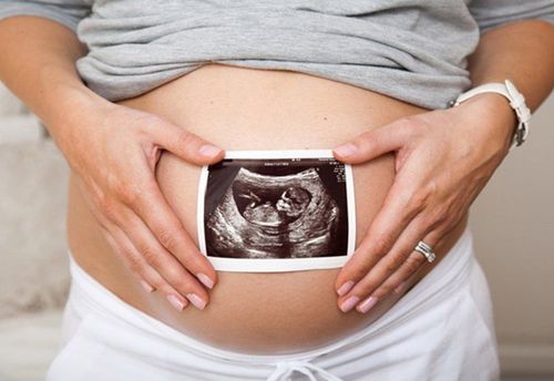
Ever since you found out you were pregnant, one of the first milestones you can look forward to is hearing your baby's heartbeat. Fetal echocardiography is also an important method in prenatal diagnosis for pregnant women.
1. When is the fetal heart?
During fetal development, the heart begins to form quite clearly and beat around 22 days after conception, often before the mother realizes she is pregnant. Fetal heartbeat will usually appear at 6-7 weeks of pregnancy, at this time with modern ultrasound techniques, we can hear the fetal heartbeat. However, in many cases, it is not until about 8 - 10 weeks of pregnancy that the fetal heartbeat can be heard, this depends on the menstrual cycle as well as the development of the embryo. During this stage, the heart develops from a simple tube shape then twists and divides, eventually forming the four chambers and heart valves (which open and close blood to release blood from the heart to all over the baby's body). From the 20th week onwards, the fetal heart beat has become strong and now just need to use normal headphones to be heard. The louder and louder the heartbeat of the fetus, the easier it is to hear, indicating that the fetus is completely healthy and developing normally.Trắc nghiệm: Bạn có hiểu đúng về dấu hiệu mang thai sớm?
Các dấu hiệu mang thai sớm không phải chỉ mỗi trễ kinh mà còn có rất nhiều dấu hiệu khác như xuất huyết âm đạo, ngực căng tức,… Điểm xem bạn biết được bao nhiêu dấu hiệu mang thai sớm thông qua bài trắc nghiệm này nhé!
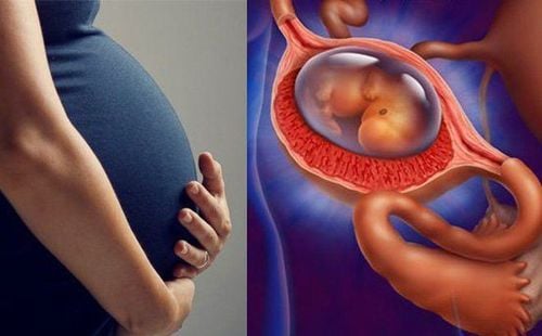
2. The process of forming fetal heart
After being fertilized in the first third of the fallopian tube, from the 30th hour, the zygote moves down to the uterus and divides exponentially 2. From the first cell, the zygote divides into 2 cells that remain attached. contiguous and then into 4, 8, 16,... After 5 days, it develops into a blastocyst. Two days later, the embryo reaches the uterus and is buried in the lining for implantation. At this stage, the embryo will secrete HCG present in the urine. Therefore, it is possible to know that you are pregnant when you try Quickstick, ultrasound at this stage may not be obvious.
Tim thai thường xuất hiện vào tuần thứ 6 - 7 của thai kỳ
During transcardiac echocardiography, cardiac activity can be determined in real-time 2D images of the uterus when the embryonic apex length is ≥ 5mm. This occurs at 5 weeks 3 days to 6 weeks 3 days of gestation, counting from the last menstrual period. After 6 weeks of gestation, spectral and color Doppler signals of pulsating blood in the fetal heart and great vessels can be detected. During the latter part of the first trimester of pregnancy, ultrasound imaging and Doppler recording can be performed transcranial.
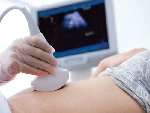
3. Time of first fetal echocardiography

Siêu âm thai 4D
During the first ultrasound, the doctor or technician will check the following:
Confirm pregnancy, check for pregnancy or ectopic pregnancy Confirm baby's heart rate Measure head length At present, Vinmec International Hospital applies 2D, 3D, 4D ultrasound techniques to help mothers hear the fetal heart from the stage of pregnancy. head. Vinmec International General Hospital system has been using the most modern generations of color ultrasound machines today in examining and treating patients. One of them is GE Healthcarecar's Logig E9 ultrasound machine with full options, HD resolution probes for clear images, accurate assessment of fetal malformations, if any. Ultrasound services are included in the Maternity Package at Vinmec.

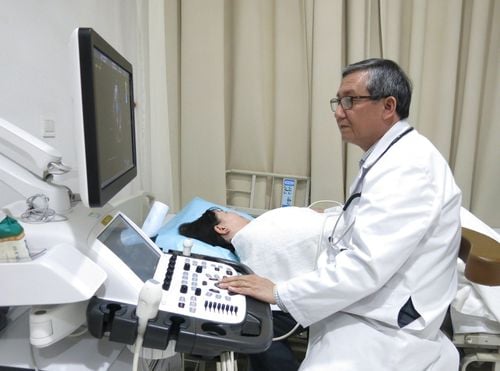
Hình ảnh khách hàng được tư vấn sức khỏe khi khám thai tại Vinmec
Please dial HOTLINE for more information or register for an appointment HERE. Download MyVinmec app to make appointments faster and to manage your bookings easily.