This is an automatically translated article.
The article is professionally consulted by Master, Doctor Nguyen Van Huong - Department of Diagnostic Imaging - Vinmec International Hospital Da Nang. The doctor has many years of experience in the field of diagnostic imaging.When hearing about the phenomenon of pregnancy ultrasound with Yolksac, many pregnant women worry about what Yolksac is? Is ultrasound dangerous Yolksac? Information about the Yolksac ultrasound phenomenon will be answered in the article below.
1. What is Yolksac Ultrasound?
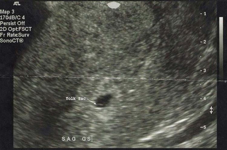
To put it simply, the yolksac is the yolk sac, formed when the fetus (zygote implanted in the uterus) is about 5 weeks old, while the embryo and fetal heart will be formed when the fetus is 6-6 years old. , 5 weeks old.
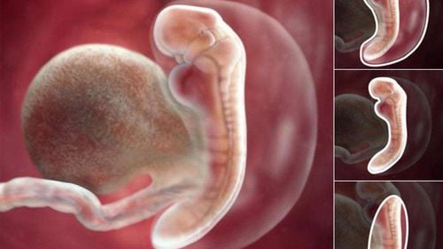
The yolk sac is the first structure formed in preparation for pregnancy. Yolksac has a role in providing nutrition to the embryo in the early stages, when the embryo has not been formed and there is no placenta. Usually, when 5 weeks pregnant ultrasound can see the yolk sac, also known as the phenomenon of ultrasound with Yolksac.
The structure of the yolk sac is formed from the endoderm of the embryo and is covered by the mesoderm visceral leaf, to form the basic cells, yolksac contains a lot of protein. During embryonic development, the endodermal cells roll back into the primitive intestinal wall and expand into the yolk sac. After the amniotic sac is formed and grows, compressing the yolksac, the yolk sac is now only a narrow tube through the middle called the yolk sac.
The size of the yolk sac is only about 5.6 mm between 5 - 10 weeks in pregnancy. As the embryo develops, the yolksac will regress and disappear. The placenta will take over the role of nurturing the fetus.
Trắc nghiệm: Bạn có biết nên khám thai lần đầu vào lúc nào không?
Việc khám thai lần đầu mang ý nghĩa rất quan trọng, giúp bạn xác định chính xác mình có mang thai hay không? Thai nhi đã vào buồng tử cung hay chưa?... Vì vậy, nếu chưa biết khám thai lần đầu vào lúc nào, trả lời nhanh 5 câu hỏi trắc nghiệm sau sẽ giúp bạn có câu trả lời.2. When is the phenomenon of ultrasound with Yolksac dangerous?
Yolksac assumes the role of providing nutrition to the fetus in the beginning as well as it will disappear on its own after a while. However, in some cases, Yolksac will be unusually thick. So when is the Yolksac size dangerous?93% not dangerous if the yolk sac is a little thicker. If the yolk sac is about 5.6 mm thick, there is no danger. However, if the yolk sac size is larger than 5.6mm, the pregnant woman is at risk of some pregnancy complications such as a high risk of miscarriage. The larger the size of the yolk sac, the higher the risk of pregnancy.
Yolksac size can be determined by conducting ultrasound. At the 10th - 12th week of pregnancy, the doctor will conduct an ultrasound to detect abnormalities in the ovarian sac, if any, and have a timely treatment plan.
To make sure the fetus develops normally, pregnant women should repeat ultrasound as prescribed by the doctor. Besides, pregnant women can perform beta - hCG test to monitor the development of the fetus.

The pregnancy process is monitored by a team of highly qualified doctors Regular check-ups, early detection of abnormalities The Package Maternity package helps to facilitate convenient for the birthing process Newborns are taken care of comprehensively.
Please dial HOTLINE for more information or register for an appointment HERE. Download MyVinmec app to make appointments faster and to manage your bookings easily.


