This is an automatically translated article.
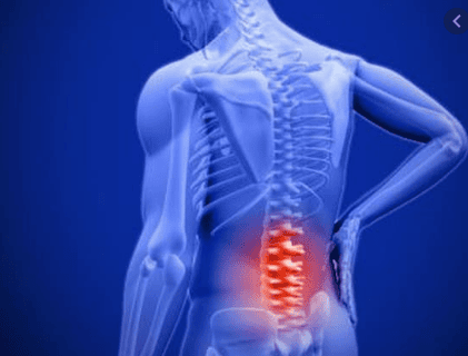
The article is professionally consulted by Master, Doctor Nguyen Le Thao Tram - Diagnostic Imaging Doctor - Imaging Department - Vinmec Nha Trang International General Hospital.X-ray of the spine is a routine imaging technique that helps doctors detect abnormalities in parts as well as the entire spine that are not visible to the naked eye.
1. Spine
The spine is the pillar that supports the entire body. The human spine usually consists of 33 vertebrae and other basic components, such as discs, spinal cord, nerve roots,...2. X-ray methods of the spine
Take X-ray of each region of the spine on both straight and inclined planes: X-ray of the cervical spine, X-ray of the lumbar spine,... Spine scans in special positions such as: C1, C2 vertebrae straight posture, open mouth; Scan 3/4 of the spine to detect foraminal changes. Contrast myelogram to detect occlusion of the canal (particularly myeloma). Radiography (especially for herniated discs in the lumbar spine). Computed tomography – Magnetic resonance imaging: is the method currently considered the most valuable in the field of medical imaging of the spine and spinal cord. Indicated in cases of suspected myeloma, disc herniation.3. What diseases can X-rays of the spine help diagnose?
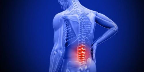
Congenital malformations of the spine Degeneration of the spine Disc herniation Spinal trauma Spinal tuberculosis Inflammatory diseases Ankylosing spondylitis Spinal cord tumor 3.1. Spinal deformities Some common spinal deformities:
Spinal deformities caused by transitional disorders: There are 8 cervical vertebrae, 6 vertebrae in the lumbar spine. Double spine and open waist: Caused by incomplete ossification process of the spine. Congenital fusion of two vertebrae: The two vertebrae are fused together both at the intervertebral disc space and at the posterior arch, the two vertebrae are not destroyed, so the spine axis is not hunched or crooked. Anterior angle ossification points exist: The ossification points of the vertebral body are located at the corners of the vertebral body. Due to incomplete ossification, the ossification point exists as a fragment of bone separated from the anterior angle of the vertebral body, usually after aseptic necrosis of the synovial cartilage (Scheuermann's disease). Scoliosis: Looking at the straight film in the anterior-posterior position, the spine is misaligned, some vertebrae are rotated with a wedge-shaped vertebral body deformity as in hemivertebral malformation. Spinal kyphosis: Looking at the lateral radiograph, the spine is protruding posteriorly, causing hunchback, due to the wedge-shaped deformed vertebrae. 3.2. Spondylolisthesis Osteoarthritis is a common disease in people over 40 years old. According to Schmorl, spondylolisthesis is degeneration of the fibrous ring surrounding the disc. As a result, the disc swells, protrudes, vertebral ligaments are stretched and calcified near the disc edge to form a bony beak. The bony beak usually appears on the anterior and bilateral sides of the vertebral body, rarely on the posterior border (if present, it will compress the spinal cord).
The height of the disc space in spondylolisthesis is little changed at first, but after a long time it is also narrowed due to osteochondrosis, especially disc degeneration.
3.3. Disc herniation Disc herniation is the herniation of the nucleus pulposus of the disc through the tear of the annulus. The migration of the mucinous nucleus is usually posterior and causes compression of the canal and nerve roots. Herniated discs are common in the lumbar spine, can occur in the cervical spine, and rarely in the thoracic spine.
Conventional radiographs do not give a direct image of disc herniation, however there is a special herniation that can show images on conventional radiographs that is herniated into the vertebral body (Schmorl hernia). This type of hernia often occurs in patients with severe spinal osteoporosis.
3.4. Spinal Injury Spinal injury is a common condition caused by traffic accidents, daily life and work. Common injuries are:
Fracture of the vertebral body: The fracture line runs across the vertebral body, causing interruption or angulation at the upper border of the vertebral body. Collapse of the vertebral body: The height of the vertebral body is flattened, the contrast density is higher than that of the adjacent vertebrae, often subsidence in the anterior border of the vertebral body, so the collapsed vertebral body has a wedge shape. The intervertebral disc space is generally not narrow. Slip of the vertebral body: Usually slides forward, backward, or to the side, which can cause pulp compression or complete severing of the pulp. Fracture of the apex C2. Transverse process fractures: Usually transverse process fractures in the bodies of the lumbar vertebrae. Posterior arch fractures, spines are uncommon.
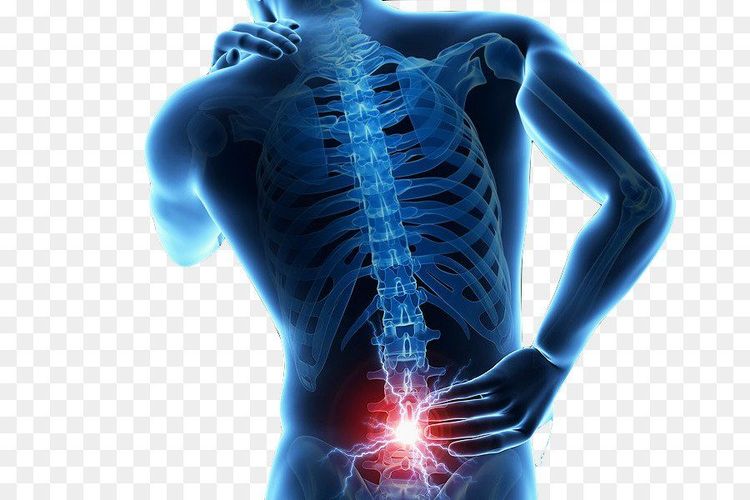
Early stage: Disc stenosis, visible on both straight and lateral films (most clearly on inclined films). In the first stage, disc stenosis is still discreet, and should be compared with the adjacent disc space. Full-blown stage: Along with signs of disc stenosis is a destructive image of bacteria entering the vertebral body. The vertebral body edge is close to the disc, the body is flattened, wedge-shaped, the physiological curve is deformed and bent forward, causing hunchback. Recovery and sequelae: Tuberculous vertebrae are fused together, disc space is lost; In the thoracic spine there is a "spider foot" shape. A cold abscess can also be seen. It is a rhombus-shaped cloud located on both sides of the affected vertebrae (usually only clearly visible in the thoracic vertebrae). Cold abscesses often appear in the full-blown stage of the disease, can cause fistulas, and persist until the tubercle is completely healed.
3.6. Ankylosing spondylitis (Bechterew - Strumpell - Marie) The beginning of Bechterew's disease is sacroiliac arthritis (accounting for 70%), followed by bone thinning in the spine. At the earliest, after 3 years, calcification of ligaments and joints of the spine will appear. When calcification of the anterior longitudinal, interspinous, and lateral ligaments of the spine will resemble the image of "bamboo spine", "train track".
Bechterew's disease is a diffuse ankylosing spondylitis, the first stage of damage is in the spine and sacroiliac joints, later a series of other joints are also glued such as the hip.
3.7. Spinal cord tumors The types of spinal tumors that can be encountered are: Epidural Tumor, Extradural Tumor, Intramedullary Tumor. For myeloma, it is also possible to use magnetic resonance imaging, which will give better results and is the most reliable imaging method in diagnosing myeloma.
Doctor Nguyen Le Thao Tram has many years of experience in the field of specialized imaging in pathology on ultrasound, radiology, multi-slice CT, magnetic resonance such as nervous system, spine, musculoskeletal system joints, soft tissue, tumor diseases, intra-abdominal inflammation and vascular diseases... In addition, Dr. Tram is also capable of performing fine needle aspiration (FNA) mammography, thyroid gland and subcutaneous soft tissue. Currently working at the Diagnostic Imaging Department of Vinmec Nha Trang International General Hospital.
Please dial HOTLINE for more information or register for an appointment HERE. Download MyVinmec app to make appointments faster and to manage your bookings easily.


