This is an automatically translated article.
The article was written by doctors of Internal Oncology Department - Department of Internal Oncology - Radiation Oncology Center, Vinmec Times City International General Hospital.1. How dangerous is kidney cancer?
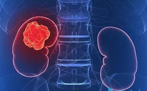
Kidney cancer is the sixth most common cancer for men and eighth for women. The disease is rare in people under 45 years of age. The median age of diagnosis is 64.The 5-year survival rate for people with kidney cancer is 75%. However, this rate depends on several factors such as the cell type and stage of the cancer when it is first diagnosed.
About two-thirds of people are diagnosed when the cancer is only in the kidney. For this group, the 5-year survival rate is 93%. If kidney cancer has spread to surrounding tissues or organs and/or regional lymph nodes, the 5-year survival rate is 69%. If the cancer has spread to a distant site of the body, the 5-year survival rate is 12%.
2. Identify signs of kidney cancer

Giảm cân không giải thích được là dấu hiệu của ung thư thận
Pain or heaviness in the side or back A mass or lump in the side or back Swelling of the ankles and legs High blood pressure Deficiency blood, low red blood cell count Fatigue Loss of appetite Unexplained weight loss Fever not related to repeated bacterial, viral, or viral infection For men, see enlarged veins around the testicles, especially the right testicle, which may be associated with a large tumor in the kidney.
3. Tests to help diagnose disease
Blood and urine tests: to check the number of red blood cells in the blood and urine tests to look for red blood cells, bacteria, or cancer cells. These tests may be suggestive, but they are not valuable for a definitive diagnosis.Biopsy: taking a small amount of tissue for examination under a microscope. Imaging tests Computed tomography Magnetic resonance imaging (MRI) Prepared urography: this test is less commonly used today because it has been replaced by CT/scan of the urinary tract, which provides an Image Clearer and deeper images of the urinary system Cystoscopy and ureteroscopy: Sometimes cystoscopy and ureteroscopy are needed for pelvic and pelvic kidney cancers. This technique can be used to take tumor cells for examination under a microscope, to perform biopsies... After these tests are done, the doctor will look at all of them to determine the stage. disease stage.
4. Diagnosing Kidney Cancer

Chẩn đoán ung thư thận như thế nào?
Tumor : T TX: Primary tumor cannot be assessed
T0 : No evidence of primary tumor.
T1: Tumor found only in kidney and largest diameter < 7 cm
T1a: Tumor found only in kidney and largest diameter < 4 cm T1b: Tumor found only in kidney and tract largest diameter between 4 cm and 7 cm T2: Tumor found only in kidney and largest diameter > 7 cm
T2a: Tumor only in kidney and largest diameter > 7 cm but not more than 10 cm
T2b: Tumor only in kidney and largest diameter > 10 cm T3: Tumor has spread into large veins in kidney or connective tissue, fatty tissue around kidney. However, the tumor has not spread to the adrenal gland wall on the same side of the body as the tumor. The adrenal glands are located on top of each kidney and produce hormones and adrenaline to help control heart rate, blood pressure, and other bodily functions. In addition, the tumor has not spread beyond the layer of tissue surrounding the kidney.
T3a: The tumor has spread to a large vein leading out of the kidney, called the renal vein, or branches of the renal vein; fatty tissue surrounding and/or inside the kidney; T3b: The tumor has grown into a large vein that drains into the heart, called the inferior vena cava, below the diaphragm. T3c: The tumor has spread to the vena cava above the diaphragm and into the right atrium of the heart or to the wall of the vena cava. T4: The tumor has spread to areas outside the renal cortex, into the adrenal glands on the same side of the body as the tumor.
Nodes (N) Lymph nodes near the kidney are called regional or regional lymph nodes. Lymph nodes in other parts of the body are called distant lymph nodes.
NX: Regional lymph nodes cannot be assessed
N0 : Cancer has not spread to regional lymph nodes.
N1: Cancer has spread to regional lymph nodes.
Distant metastasis (M) Common areas where kidney cancer can spread include the bones,
liver, lungs, brain, and distant lymph nodes.
M0 : The disease has not spread far away
M1: The cancer has spread to other parts of the body other than the kidney.
Staging Combining the T, N, and M classifications gives the stage
Stage I: T1, N0, M0.
Stage II: T2, N0, M0
Stage III: One of the following conditions:
T1 or T2, N1, M0. T3, any N, M0 Stage IV: One of the following:
T4, any N, M0 Any T, any N, M1. Recurrence: Recurrent cancer is cancer that has come back after treatment. Lesions may be found in the kidney area or in another part of the body. If the cancer comes back, there are a number of other tests to find out about the extent of recurrence. These tests are often similar to those performed at the time of initial diagnosis.
Please dial HOTLINE for more information or register for an appointment HERE. Download MyVinmec app to make appointments faster and to manage your bookings easily.
Source Cancer.net 2019


