This is an automatically translated article.
The article is professionally consulted by Master, Doctor Lam Thi Kim Chi - Department of Diagnostic Imaging - Vinmec International General Hospital Da Nang.Magnetic resonance is not only the most modern fast and accurate imaging method today, but also plays a very important role in the examination and treatment of prostate cancer in men.
1. The role of magnetic resonance imaging in prostate cancer diagnosis
Prostate cancer is one of the most common cancers in men, the second leading cause of cancer death in men (after lung cancer). Accordingly, when prostate cancer is still in the capsule, the survival rate of the patient after 5 years is up to 100%. Therefore, when the disease is diagnosed early, the chance of cure is very high.
Prostate magnetic resonance imaging using contrast is the method of choice for many large hospitals (including Vinmec International General Hospital) because of its high resolution and soft tissue contrast. Multi-faceted, easy to assess prostate cancer stage before treatment and follow up after treatment. In addition, functional and dynamic magnetic resonance with contrast agents improve the sensitivity and specificity in diagnosis

Chụp cộng hưởng từ giúp phát hiện sớm và chính xác ung thư tuyến tiền liệt
2. Indications and contraindications of magnetic resonance imaging for prostate cancer treatment
Indications for subjects:
All cases of male patients with suspected prostate cancer based on clinical diagnosis or imaging and other tests. Evaluation of complications after pelvic surgery Before biopsy for cancer Evaluation after radiation and chemotherapy Prostate infection or abscess Diagnosis of recurrent prostate cancer Preoperative assessment and any abnormalities Usually congenital prostatic hypertrophy Contraindicated for the following subjects:
Prostate magnetic resonance imaging is indicated and contraindicated in the following cases:
Absolute contraindication
Patients in During the scanning process, electronic devices such as pacemakers, anti-vibration machines, hearing aids, cochlear implants, metal jewelry, automatic subcutaneous drug injection devices, Neurostimulator, etc. must not be carried. Metal clips used in intracranial, orbital, vascular surgery under 6 months. If the patient's condition is severe, resuscitation equipment is needed next to the person. Relative contraindications
Metal surgical clamps over 6 months Patient's psychology such as: Fear of the dark, fear of narrow spaces, fear of being alone,...
3. Prepare the patient before the scan
Drugs used during magnetic resonance imaging:
Sedatives Magnetic contrast agents Antiseptics for skin and mucous membranes. Notes for the patient:
It is necessary to listen to and follow the instructions of the medical staff in charge of the scan to make the working process go smoothly. Observe the contraindications mentioned in section 2. Use special clothing for the magnetic resonance imaging room. There is a request for a scan from a doctor who makes a clinical diagnosis and complete medical records (if necessary). Have the patient hold urine for 15-20 minutes before the exam The procedure is explained Is given earplugs or ear protection Remove anything that contains metal (dentures, hearing aids, hairpins, pages, etc.) pressure, ear loops...) Place an intravenous line before entering the imaging room Make sure that the patient understands and fills in the consent form for magnetic resonance imaging.

Bệnh nhân cần tuân theo hướng dẫn của cán bộ y tế trong quá trình chụp cộng hưởng từ
4. Steps to conduct prostate resonance imaging
In order for magnetic resonance imaging to bring high results, the patient needs to follow the exact steps required by the doctors and nurses:
Patient's lying position
The doctor instructs the supine position magnetic resonance imaging table. Select the position and position the receiver coil. Move the table into the magnetic field of the machine and locate the imaging area Prostate magnetic resonance imaging technique
Place an intravenous line with an 18G needle, connected to a double-bore electric injection pump, one of which contains the drug magnetic contrast medium and a barrel containing physiological saline. The usual amount of contrast agent used is 0.2ml/kg body weight. Localization Imaging before contrast injection
Pulse sequence 1: Transverse T2W, above the pelvic floor (instructions on image showing the vertical plane), slice thickness 3-4 mm, distance between slices 10% of the slice thickness (0.3-0.4 mm or a factor of 1.1) Pulse sequence 2: Transverse T1W transverse section of the prostate (vertical profile), layer thickness cut 2-3 mm, increments of 0-10% of cut thickness (0-0.3 mm or scale of 1.0-1.1) 3rd pulse sequence: horizontal T2W, 3 mm of cut thickness, 0-10 increments % of cut thickness (0-0.3mm or 1.0-1.1 ratio) 4th pulse sequence: Cross sectional T2W, 3mm cut thickness, 0-10% increments of cut thickness (0-0.3mm or ratio 1.0-1.1) Taken after injecting magnetic contrast Contrast was injected with 0.1 mmol gadolinium/kg body weight, at a rate of 2ml/sec. 5th pulse sequence: Transverse T1W, similar to 2nd sequence: 6th pulse sequence: cross-section of the prostate in vertical or horizontal plane, layer thickness of 2-3 mm, increments of 0-10% of surface thickness of the cut (0-0.3mm or 1.0-1.1 scale). For patients with cause, it is possible to use an intrarectal coil or an abdominal wall coil located on the abdomen in the pelvic region and fixed with a lifeline, using a small field of view.
5. Evaluation after magnetic resonance imaging
Magnetic resonance imaging must clearly show the entire prostate gland and adjacent organs such as seminal vesicles, rectum ... in the vertical, horizontal and horizontal directions. Assess the extent of contrast enhancement of the lesion (if any).
6. Complications during the shooting process and how to handle it
When performing magnetic resonance imaging technique, if an accident occurs, the doctor should quickly handle the accident as follows:
Patient's state of fear, agitation, psychological panic. At this time, the doctor performing the scan needs to encourage, comfort and reassure the patient's spirit. In case of anxiety and fear, sedation can be used under the supervision of an anesthesiologist. Complications related to contrast agents are handled according to the issued regulations and guidelines of the Ministry of Health. Master Doctor Lam Thi Kim Chi graduated with a Master's degree in Radiology - Hue University of Medicine and Pharmacy and has over 6 years of experience as a radiologist. Doctor Chi used to work at Da Nang Obstetrics and Children's Hospital before becoming a radiologist at Vinmec Danang International Hospital as it is today
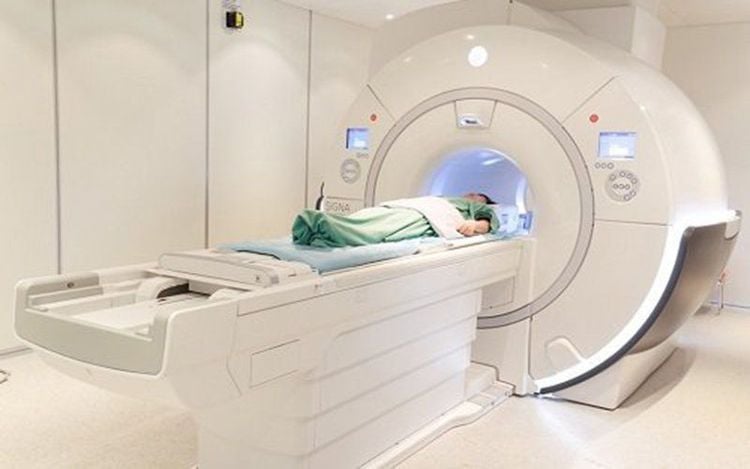
Currently Vinmec International Hospital is a single the first to put into use a 3.0 Tesla magnetic resonance imaging machine with Silent technology, bringing outstanding advantages.
To schedule an MRI scan at Vinmec with today's most modern magnetic resonance machine system, you can immediately contact the nearest Vinmec International General Hospital facility system, or register for a medical examination. online HERE.
MORE:
MRI scan – the "golden" method to accurately diagnose disc herniation Bone scintigraphy detects early-stage bone metastatic cancer and other bone lesions First time using magnetic resonance imaging “no noise” at Vinmec Nha Trang


