This is an automatically translated article.
Radiation therapy is an almost indispensable method of cancer treatment, but today, in addition to conventional radiation therapy, there are many more advanced methods of radiation therapy, including radiation therapy with a linear accelerator.
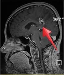
1.What is linear accelerator SRS radiosurgery? Scientific name: SRS radiosurgery using linear accelerators Preliminary technical description: SRS brain radiosurgery is a method of using multiple beams of radiation that are not too strong. The radiation receivers concentrated in the brain tumor will receive a very large dose of radiation to kill the cancer cells. SRS brain radiosurgery is usually done in one treatment, and doctors can destroy multiple tumors in one treatment.
Most cases can go home the same day. The risk of long-term cognitive decline after SRS treatment is believed to be less than with whole brain radiation, increasing the patient's chances of survival, cognitive ability, and quality of life.
2.Indications and contraindications Indications:
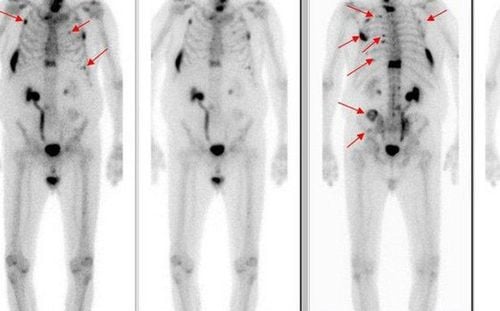
Patients are diagnosed with cancer, have brain metastases 1-3 foci with the largest diameter ≤ 4cm, good health, prognosis for survival ≥ 3 months.
3.Advantages and disadvantages of the technique Advantages
The technique helps only 1-3 sessions of radiation, the patient does not have to be hospitalized, most importantly preserves memory, can re-radiate the brain to other locations, whole brain radiation if there is diffuse progression after radiation > 3 months.
Disadvantages
Limitations in the treatment of small lesions in the brain region, such as arteriovenous malformations (AVMs), auditory neuromas

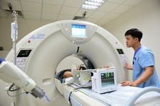
Xạ phẫu SRS bằng máy gia tốc tuyến tính là kỹ thuật cao đòi hỏi có chuyên gia nhiều kinh nghiệm
4. Procedure Step 1: Put the patient in the patient's position
The patient lies on his or her back, arms stretched along the body. Step 2: Immobilize the patient
Immobilize the head with a suitable set of pillows and an Encompass kit (Qfix) or a head and neck mask (Qfix/Civco), which can be combined with a head and neck vaccine. Immobilize legs and arms using Shoulder retractor. Step 3: Simulation, image data collection for planning
CT scan simulates contrast injection 3 series of images: before contrast injection, artery and portal vein with contrast injection, slice cut 2.5mm, step 2.5mm from apex of lung to iliac crest. In addition, it is possible to take additional series of images for drawing beer volume. Step 4: Perform patient placement, centralization, EPID verification and irradiation
Must be present Doctor, Technician, Engineer Pre-irradiation image verification is done daily, right before irradiation : Normally 01 verification will take 02 films: 01 film in AP direction, 01 film in LAT direction. Take EPID and centripetal fluid: kV-CBCT, if metal instruments are available, use MV-Cone Beam CT. Perform the treatment center shift procedure to ensure that the setting error is only ≤ 1 mm. Carry out irradiation. 5. Normal manifestations after the technique. Patients may have symptoms depending on the size and location of the tumor.
Fatigue, anorexia, vomiting, nausea (when chemotherapy and radiotherapy are concurrent) Radiation dermatitis Radiation pneumonia (chest radiotherapy) 6.When are the following symptoms when performing the technique? often and in need of immediate re-examination? Skin atrophy, skin necrosis in radiation treatment area Dry mouth, tight jaw (radiotherapy to the head and neck area) Pulmonary fibrosis (radiotherapy to the chest area) 7. Types of machines/equipment required to perform this technique Radiotherapy machine Truebeam speed NDS120HD V2.7
Please dial HOTLINE for more information or register for an appointment HERE. Download MyVinmec app to make appointments faster and to manage your bookings easily.