This is an automatically translated article.
This article is professionally consulted by Master, Doctor Vu Thi Hau - Radiologist - Department of Diagnostic Imaging and Nuclear Medicine - Vinmec Times City International General Hospital. The doctor has many years of experience in the field of diagnostic imaging including magnetic resonance, CT and ultrasound.X-rays help monitor the progress of emphysema in the body that cannot be seen with the naked eye. X-rays are used to diagnose emphysema more accurately.
1. What is emphysema?
Emphysema occurs when the alveoli are stretched so often that they lose their elasticity and are no longer elastic; Gas entering the alveoli is stuck and difficult to escape, causing gas stagnation in the alveoli and reducing the ability to exchange O2 and Co2.2. Causes of emphysema
2.1 Smoking or inhaling tobacco smoke regularly This is the leading cause of disease. Because in cigarettes there are more than 4,000 toxic chemicals that stimulate the destruction of small peripheral airways, air sacs, elasticity and the support of elastic fibers.2.2 Protein deficiency This case accounts for only 1-2% of people with emphysema due to hereditary deficiency of AAT protein, which protects the elastic lung structure. Without this protein, enzymes cause progressive lung damage, eventually causing emphysema.
2.2 Occupational characteristics The trumpeters and glass players have a high risk of emphysema because their occupation can cause alveolar dilatation and increased intra-alveolar pressure.
3. Classification of emphysema
3.1 Primary emphysema
Central cystic or central lobular emphysema is also called type B, or blue or purple edema. Distal alveolar emphysema 3.2 Secondary emphysema Emphysema around bronchiole function or focal and adjacent to fibrous tissue.
4.Symptoms of emphysema
Shortness of breath, pursed lips or wheezing Chest tightness, shortness of breath Cough, possibly due to mucus secretions Mobility and activity diminished over time, as the disease develops slowly Feeling tired, loss of appetite, weight loss5. Diagnosis of emphysema
5.1. Tests to diagnose emphysema To determine exactly, the doctor may order certain tests, specifically:Spirometry and pulmonary function tests (PFTs) Blood gas analysis artery Pulse oximetry method Sputum examination: Analysis of sputum cells and computed tomography (CT scan) X-ray 5.2. The role of X-ray in the diagnosis of emphysema

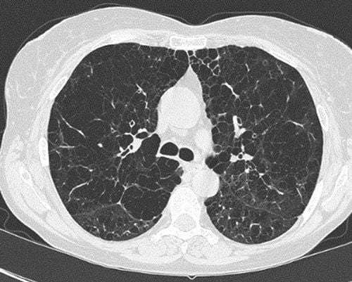
Diagnosis of emphysema through X-ray film:
X-ray film shows: The lungs are bright, the intercostal space is dilated, the diaphragm is lowered and breathing movements are reduced. X-ray films can diagnose 3 main symptoms of the lungs: Stretching of the lungs; reduced pulmonary circulation, emphysema. The pathology of emphysema can be identified when seeing: Dirty image in the lungs, dilated lung syndrome. The nodules on both sides of the thorax, along with the blood vessels of the upper lobe, are sparse. The arteries in the lungs are dilated. Inflammation around the bronchioles. Emphysema X-ray is a true basis for determining internal changes in the lungs and monitoring the evolution of emphysema in the human body. However, correct diagnosis is only the first step of success, from diagnosing emphysema through X-rays, doctors and patients need the best treatment regimen to overcome the condition of the disease.
Please dial HOTLINE for more information or register for an appointment HERE. Download MyVinmec app to make appointments faster and to manage your bookings easily.


