This is an automatically translated article.
The article was professionally consulted by Dr. Nguyen Anh Tu - Doctor of Obstetric Ultrasound - Prenatal Diagnosis - Obstetrics Department - Vinmec Hai Phong International General Hospital.The article was professionally consulted by Specialist Doctor I Le Hong Lien - Department of Obstetrics and Gynecology - Vinmec Central Park International General Hospital. Doctor Lien has over 10 years of experience as a radiologist in the Department of Ultrasound at the leading hospital in the field of obstetrics and gynecology in the South - Tu Du Hospital.
Fetal echocardiography is a medical imaging method to assess the cardiovascular status of the fetus such as: heart rate, fetal heart function... Since then, it can detect severe heart defects early, helping to intervene promptly. treatment from the very beginning of pregnancy.
1. What week does the fetal heart appear?
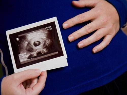
During fetal development, the heart begins to form quite clearly and beat around 22 days after conception, often before the mother realizes she is pregnant. Fetal heartbeat usually appears in the 6-7 week of pregnancy, at this time with modern ultrasound techniques can hear the fetal heartbeat. However, in many cases it is not until about 8 to 10 weeks of pregnancy that a fetal heartbeat can be heard. This also depends on the menstrual cycle (the calculation of gestational age is based on the menstrual cycle) as well as the development of the embryo.In the early stages, the heart develops from a simple tube shape then twists and divides, eventually forming a heart with four chambers and valves. From the 20th week onwards, the fetal heartbeat has become strong and now just need to use the ear to be able to hear it. If the heartbeat of the fetus is louder and easier to hear, it means that the fetus is very healthy and develops normally.

2. Fetal echocardiography for what?
According to the latest statistics of the Ministry of Health, each year in our country, about 9,000 - 10,000 children are born with congenital heart disease, this rate accounts for 0.8% of children born in the same year. Among them, 50% of children with very severe congenital heart disease and only half of children with congenital heart disease have surgery, the rest have to live with the disease and face the risk every day. However, checking for congenital heart defects is something that pregnant women often miss. Fetal echocardiography is a medical imaging method performed by doctors with extensive training in fetal cardiology.Fetal echocardiography helps support doctors in assessing fetal cardiovascular status such as: heart rate, fetal heart function, congenital heart defects. Doctors also recommend that this method should be included in prenatal diagnosis to help detect severe heart defects early, helping to timely intervene in treatment right from the early stages of pregnancy.
Currently, fetal echocardiography has made great strides in technology. Fetal echocardiography can detect about 60% of fetal heart abnormalities. Ultrasound consultation after screening, then diagnosis will have accurate results about 90% of antenatal fetal heart diseases. Although during this period, the fetal heart is a structure that develops and changes day by day.
3. Which pregnant women should perform fetal echocardiography?
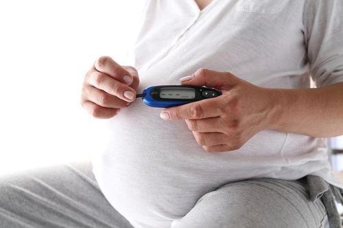
Most babies born with congenital heart disease have no prior signs of risk. Therefore, obstetricians and gynecologists recommend all pregnant women to perform fetal echocardiography during pregnancy. Special attention should be paid to pregnant women in high-risk groups such as:Abnormalities were detected in routine pregnancy ultrasound. Using drugs that affect the fetus such as: anticonvulsants, antidepressants, prostaglandin synthesis inhibitors (salicylic acid, ibuprofen, indomethacin...) ... The fetus is artificially inseminated. There is a family history of congenital heart disease. A mother who has had a child with a congenital heart defect has a 1/20 - 1/100 chance of having the disease. If the previous two children had congenital heart defects, the fetus has a risk of 1/10 - 1/20. In case the mother has a congenital heart disease, the fetus has a risk of 1/5 - 1/20. If the father has the disease, the baby has a 1/30 chance of having the disease, which means the baby has a 3% chance of getting the disease. Pregnant women with diabetes, Phenyl ketones in urine or some other genetic diseases such as: Ellis Van Creveld, Marfan, Noonan... Pregnant women with Rubella infection, autoimmune diseases (lupus red, Sjogren's HC...) during pregnancy.

4. How should the fetus need to perform fetal echocardiography?
The method of fetal echocardiography is also indicated by doctors for fetuses in the group that are prone to congenital heart disease:Fetal heart arrhythmia. Suspected twin exchange syndrome or multiple pregnancies. The fetal nuchal translucency increases during the first 3 months of pregnancy. Placental edema is not hereditary. Extracardiac abnormalities. Chromosomal abnormalities: umbilical hernia, nuchal edema, duodenal bulb atrophy, diaphragmatic hernia,... Usually fetal echocardiography will be performed around 18-24 weeks of pregnancy. However, pregnant women should pay attention to carefully check the fetal heart during the whole pregnancy and the most important milestone is 22 weeks. Currently, fetal echocardiography to detect malformations tends to be performed as early as 16-18 weeks gestation, as early as 12-13 weeks gestation. From 12 weeks of age, the baby has developed relatively fully. in terms of morphology and has reflexes such as flexing and stretching the body, stretching the limbs... This is also one of the 03 important ultrasound landmarks recommended by experts to perform. During this ultrasound, doctors will especially check and screen for early abnormalities of the brain, face, heart, digestive, urinary, extremities and the whole body. Because the fetus is still quite small, a high-end 4D ultrasound system like the GE Voluson E10 will play a very important role in helping doctors at Vinmec detect more than 95% of anomalies during this period. Packed with the latest technologies, the GE Voluson E10 enables enhanced image quality and penetration for outstanding high-resolution images and easy operation. However, fetal echocardiography at 22 weeks is still the most important.
Currently, all Maternity packages at Vinmec International General Hospital have fetal ultrasound, which helps to detect abnormal problems during pregnancy, and protects the health of mother and baby. Depending on the stage of pregnancy, pregnant women will be consulted by the doctor for appropriate fetal ultrasound to check the development status of the baby.
Maternity packages at Vinmec International General Hospital include:
Maternity care program 2019 – Labor Maternity care program 2019 – 36 weeks Maternity care program 2019 – 27 weeks Maternity care program 2019 – 12 weeks For more information, please contact the hospitals and clinics of Vinmec Health system nationwide.
Please dial HOTLINE for more information or register for an appointment HERE. Download MyVinmec app to make appointments faster and to manage your bookings easily.


