This is an automatically translated article.
The article is professionally consulted by Master, Doctor Ton Nu Tra My - Department of Diagnostic Imaging - Vinmec Central Park International General Hospital.X-rays of the uterus and fallopian tubes are used to see if the fallopian tubes are (partially or completely) blocked and to see if the shape and size of the uterus is normal.
1. What is an X-ray of the uterus and fallopian tubes?
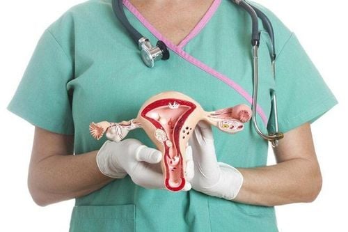
An X-ray of the uterus and fallopian tubes (referred to as an HSG) is an imaging test used to view the ovaries and fallopian tubes (fallopian tubes, fallopian tubes).When performing this technique, it is necessary to inject contrast material into the uterine cavity, through the vagina and cervix. When the uterus is filled with contrast fluid and the fallopian tubes are open, contrast fluid fills the fallopian tubes.
2. What is X-ray of the uterus and fallopian tubes for?
X-rays of the uterus and fallopian tubes will help with some causes of infertility and problems with pregnancy. An X-ray of the uterus and fallopian tubes is also used a few months after the tubal ligation procedure to make sure the fallopian tubes are completely blocked.However, X-rays of the uterus and fallopian tubes are not performed for the following:
Pregnant women; Pelvic infection; Heavy uterine bleeding at the time of the procedure.

3. Limitations of X-ray of the uterus and fallopian tubes
Some limitations of X-rays of the uterus and fallopian tubesAn X-ray of the uterus and fallopian tubes only helps to see the inside of the uterus and fallopian tubes. To evaluate abnormalities in the ovaries, uterine abnormalities and other pelvic structures, the patient needs to perform additional techniques such as: Uterine ultrasound, ovarian MRI, and MRI. uterus... Because infertility problems due to other causes often cannot be assessed by X-ray techniques of the uterus and fallopian tubes alone. In order to evaluate the abnormal status of the ovaries, pregnancy as well as accurately diagnose infertility in addition to the X-ray of the uterus and fallopian tubes, it is necessary to combine with some other diagnostic measures such as:
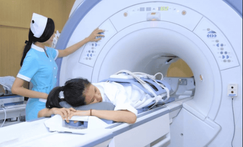
Ultrasound of the ovaries Eggs: Helps to clearly observe the status of the ovaries, evaluate abnormalities in the ovaries and uterus, monitor tumors, uterine fibroids, ovarian cysts and other gynecological diseases. Ovarian MRI, uterine MRI: Identify congenital abnormalities, detect and stage gynecological malignancies (uterus, cervix, vulva, fallopian tubes, ovaries), identify complicated adnexitis or ovarian cyst, pyelonephritis, suspect endometriosis, determine the number and location of uterine smooth muscle tumors before performing surgery to remove the tumor, hysterectomy...
4. Procedure for X-ray of uterus and fallopian tubes
4.1. Preparation before the procedure X-ray of the uterus and fallopian tubes should be done at 7-10 days from the first day of menstruation, but need to ensure the time before ovulation to be sure. The patient was not pregnant during the procedure. Inform the doctor immediately if the patient is pregnant.
The patient needs to report to the doctor about the history of drug allergies, if any, especially to the iodinated contrast medium, and sign the consent form for the procedure. The patient then needs to change clothes, put on a hospital gown, and go to the bathroom to empty the bladder.
4.2. Procedure Steps This procedure is similar to a gynecological examination. You will lie on your back on the imaging table with your knees bent, or your feet bent. The doctor will insert the speculum into the vagina, clean the cervix and insert a small catheter specifically for hysterosalpingograms into the cervix, then remove the speculum and carefully place you under the X-ray machine. The doctor injects contrast material into the uterine cavity and fallopian tubes through the catheter, then takes the necessary images. Your doctor may ask you to change positions during the scan.
After checking that the obtained images are sufficient for diagnosis, the doctor will remove the catheter and instruct you on some post-scan problems.
This method usually lasts about 30 minutes.
4.3. After the procedure The patient should use tampons after the X-ray of the uterus and fallopian tubes, however, tampons should not be used.

5. How long does an X-ray of the uterus and fallopian tubes take?
X-rays of the uterus and fallopian tubes are usually done within 30 minutes, and X-rays of the uterus and fallopian tubes can be performed as an outpatient (no hospitalization required) in a hospital's imaging department.Please dial HOTLINE for more information or register for an appointment HERE. Download MyVinmec app to make appointments faster and to manage your bookings easily.


