This is an automatically translated article.
The article is professionally consulted by Master, Doctor Nguyen Huy Nhat - Department of Medical Examination & Internal Medicine - Vinmec Danang International General Hospital.
Pulmonary function testing is a test of respiratory function that measures how well you are taking in air, breathing air out of your lungs, and how much oxygen is entering your body. The most common pulmonary function measurements are spirometry, diffusion studies, and spirometry. So what is the meaning of the respiratory function indicators?
1. Measure respiratory function for what?
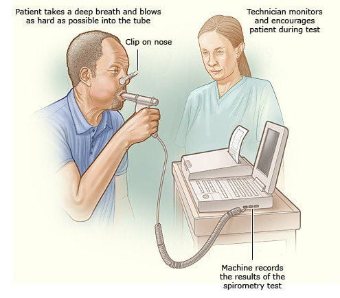
Pulmonary function measurement is a technique commonly used in the diagnosis and monitoring of the severity of respiratory diseases. The technique helps to record respiratory parameters related to lung activity, thereby helping to evaluate two syndromes of ventilation disorders: Obstruction and restriction.Meaning of the respiratory function indices gives accurate information about the air flow in the bronchi and lungs, and allows the assessment of the degree of bronchial obstruction and the severity of the dilation alveoli.
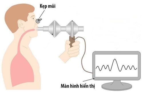
Respiratory function measurement results are expressed in specific numbers and as a percentage of the value of a normal person. The measured values of respiratory function are then plotted as a curve in which one axis represents the tidal volume measurements, the other represents the measurements of the available air volumes. in the lung, this curve is also known as the volume flow curve.
Measurement of respiratory function is a fairly simple and painless test for the patient, with almost no discomfort or complications. The accuracy of the pulmonary function test results depends on the cooperation of the patient.
2. Meaning of respiratory function indicators
2.1. Respiratory volumes
TV: The tidal volume during a normal inhalation or exhalation, in an adult normal tidal volume is about 500ml; IRV: Inspiratory reserve volume is the extra volume of air inhaled after a normal inspiration. This volume in a normal person is about 1500-2000ml, accounting for 56% of vital capacity; ERV: Expiratory reserve volume is the maximum expiratory volume after a normal expiration. This volume in a normal person is about 1100-1500 ml, accounting for 32% of vital capacity; RV: Volume of residual gas measured according to the principle of gas dilution (nitrogen or helium). Normally, the volume of residual gas is about 1000-1200 ml.2.2. Respiratory volumes

2.3. Breathing flows
\Respiratory flow is the volume of air that is mobilized in a unit of time (liters/minute or liters/second), which represents the ability or rate of gas mobilization to meet body needs and airway clearance. gas conduction.Measurement of vital vital capacity and analysis of FVC graph over time will show parameters of interval flow, point flow.
Expiratory flows include:
First Second Forced Expiratory Volume Flow (FEV1): This is the volume of air you can blow out during the first one second of expiration. Normally you can usually blow most of the air out of your lungs within a second;
Peak flow (PEF): Is the flow out of the lungs during maximal expiration, at the beginning of expiration it depends on the force produced by the expiratory muscle and the diameter of the airway, that is on exertion, then independent of exertion;
Currently, there are different types of instruments for measuring PEF. The patient inhales deeply then exhales forcefully into the device, the number indicated on the device to which the needle is pointing is the maximum expiratory flow. The difficulty may be that the patient cannot inhale deeply or exhale with maximum force or exhaled air. PEF decreases when the airways are narrowed (asthma, chronic obstructive pulmonary disease, tumor in the upper airway) or the expiratory muscles are weak.
Alveolar Ventilation Flow: The level of air exchanged in all alveoli in one minute, the effective ventilation rate. Exhaled air is a mixture of air contained in alveoli that exchange gases with blood, and air contained in the airways that do not exchange gases with blood (called the "dead space" of the respiratory apparatus). . Dead spaces include:
+ Anatomical dead space: The space in the respiratory apparatus that does not have a gas exchange area with the blood, including all the airways;
+ Physiological dead space: The anatomical dead space plus the alveoli that cannot exchange gases with the blood (such as alveolar fibrosis, capillary and alveolar constriction...);
+ The volume of air in the dead space is always changing because the airways of the respiratory apparatus are not rigid tubes, averaging about 140 ml;
+ Deep breathing is more beneficial than shallow breathing because breathing slowly and deeply reduces dead space, increases alveolar ventilation, and increases gas exchange efficiency (sustained method).

3. Respiratory function measurement results
A pulmonary function test result will usually look like this:Normal; Obstructive syndrome; Restrictive syndrome; Combination of obstructive/restrictive syndrome. A normal pulmonary function test result will vary depending on age, physical condition, race, and sex. The normal range is shown on a chart and is referred to by your doctor as he or she evaluates your measurements.
If you have a need for consultation and examination at Vinmec Hospitals under the nationwide health system, please book an appointment on the website for service.
Please dial HOTLINE for more information or register for an appointment HERE. Download MyVinmec app to make appointments faster and to manage your bookings easily.


