This is an automatically translated article.
Article written by Dr. Nguyen Quang Nam, Department of Obstetrics and Gynecology, Vinmec Times City International General Hospital
Hysterosalpingograms are used in diagnosing causes of infertility and pregnancy problems such as blocked fallopian tubes and abnormal uterine size and shape. However, not everyone can use this imaging method. So how is the process done, is there any risk?
1. What is a hysterosalpingogram (HSG)?
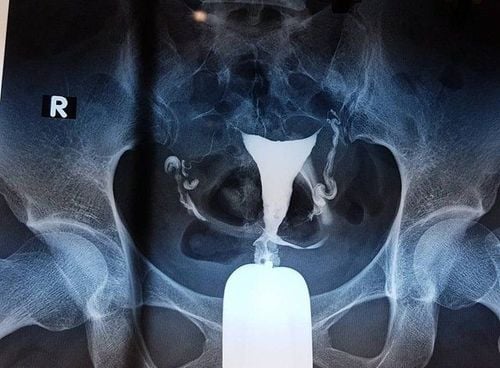
Hysterosalpingography (HSG) is a very common test in uterine and fallopian tubes where X-rays are used to view the inside of the uterus and fallopian tubes. HSG is often used to see if the fallopian tubes are partially or completely blocked, and can also show if the inside of the uterus is a normal size and shape. All of these problems can lead to infertility and problems with pregnancy.
HSG is not performed if a woman has any of the following conditions:
Pregnancy; Pelvic infection; Heavy uterine bleeding at the time of the procedure.

2. What to do to prepare for HSG hysterosalpingogram?
After your period is clear, you can have an HSG scan, in most cases an HSG is painless.
In some cases, they may also prescribe antibiotics for you prior to the HSG. Most people are able to drive themselves home after an HSG. However, you may not feel well after the procedure, so you can arrange for someone to drive you home.
3. How is HSG done?
HSG is done at the hospital. It is best to do an HSG during the first half (days 1–14) of your menstrual cycle. This period reduces the chances of pregnancy.
During HSG, a little contrast agent is injected in the uterus and fallopian tubes. This is a contrast liquid. The drug shows up in contrast to body structures on X-ray films. Contrast outlines the size and shape of the inside of the uterus and fallopian tubes. You can also see how the drug moves through the uterus and fallopian tubes.
The procedure is done as follows:
You will be asked to lie on your back with your feet flat like in a gynecological exam. A device called a speculum is inserted into the vagina. It keeps the walls of the vagina apart so that the cervix can be seen. The cervix is cleaned. An instrument called a catheter is inserted into the cervix to inject contrast material into the uterine cavity.

The speculum is removed and you are placed under the x-ray machine. The fluid is slowly introduced through the tube or ducts into the uterus and fallopian tubes. X-ray images are taken as the contrast agent fills the uterus and fallopian tubes. You may be asked to change your location. If there is no blockage, the liquid will slowly overflow to the distal ends of the tube. After it spills out, the liquid is absorbed by the body. Notes after HSG: After HSG, you may have sticky vaginal discharge because some of the fluid that comes out of the uterus is a contrast medium so you don't need to worry. The fluid may be tinged with blood. It is possible to use tampons without using tampons.You should contact the hospital if you have the following symptoms:
Vaginal bleeding; Cramp; Feeling dizzy, faint, or stomach upset.
4. What are the risks associated with HSG?
Serious problems after HSG are rare. These include an allergic reaction to the contrast medium, damage to the uterus, or a pelvic infection. The following symptoms may be present:
Vaginal discharge with a foul odor; Vomiting; Fainting; severe abdominal pain or cramps; Heavy vaginal bleeding; Fever or chills.

5. Are there alternatives to HSG?
Laparoscopy : Laparoscopy to explore the uterus and fallopian tubes is the gold standard to evaluate whether the fallopian tubes are blocked or not, but this is an invasive procedure, complicated procedure and high cost. hysteroscopy: this process can give a detailed view of the inside of the uterus. However, it cannot tell if the fallopian tubes are blocked. Uterine ultrasound: 2D ultrasound alone, or a combination of intrauterine infusion, 3D ultrasound are techniques that use ultrasound waves to display the inside of the uterus. Like hysteroscopy, it does not give us information about tubal patency or blockage. Hycosy: An ultrasound technique combined with injection of liquid containing microfoam can safely provide information about the uterus and fallopian tubes. The disadvantage of this method is its high cost and subjective dependence on the sonographer. The results of an X-ray of the uterus and fallopian tubes depend a lot on the expertise of the technicians and the equipment. Therefore, you should go to a reputable medical facility. Vinmec International General Hospital is highly appreciated for the quality of imaging techniques, including X-ray of the uterus and fallopian tubes.
A team of well-trained, professional, highly qualified and experienced technicians and doctors, regularly updated knowledge about new and advanced imaging techniques in the world world, helping to make the most accurate diagnosis for patients.
Modern equipment, completely imported from the UK, France, USA, Singapore, Japan, Korea, brings clear images and accurate results.
After the scan results are available, if there is a medical condition such as infertility, the patient will be consulted by a fertility specialist for the best treatment. Vinmec IVF Reproductive Center is the address of infertility treatment - infertility is chosen by many couples. So far, the Center has performed fertility support for over 1000 infertile couples with a success rate of over 40%. This rate is equivalent to developed countries such as UK, USA, Australia.
Please dial HOTLINE for more information or register for an appointment HERE. Download MyVinmec app to make appointments faster and to manage your bookings easily.


