This is an automatically translated article.
Fetal doppler ultrasound is a fairly common ultrasound technique that helps doctors check, diagnose and assess the health of the fetus. Fetal doppler ultrasound indicators allow survey of fetal size, hemodynamic status such as umbilical artery, cerebral artery,...
1. Fetal doppler ultrasound
Fetal doppler ultrasound is one of the most popular ultrasound techniques today. Fetal doppler ultrasound helps to measure blood flow in different locations of the fetus such as the heart, brain, umbilical cord,... to check if the baby is getting enough oxygen and nutrients through the placenta. This is the difference that conventional ultrasound cannot check.
Fetal doppler ultrasound technique is considered safe and poses no danger to the fetus and mother if performed with the frequency of ultrasound as prescribed by the doctor in the last 3 months of pregnancy. However, not all pregnant women need to perform fetal doppler ultrasound, this ultrasound technique is indicated for cases of fetal growth retardation, Rh incompatibility, low amniotic fluid index or identical twins. eggs, multiple pregnancy, abnormal mother's weight,...
MORE: Indication of Doppler ultrasound 32 weeks pregnant
2. Indicators in fetal doppler ultrasound
2.1 Fetal size
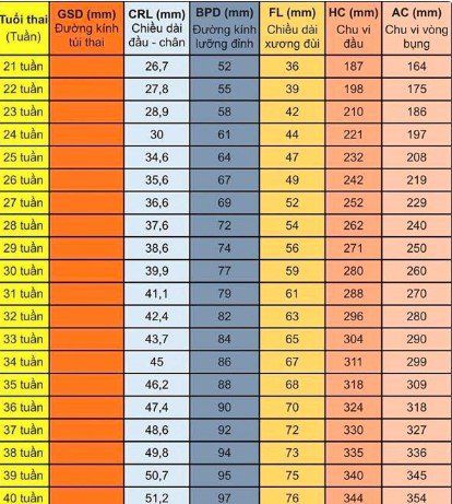
Estimation of fetal weight by clinical and ultrasound will have a decisive role in the management of poor fetal growth. Through multiple ultrasounds, the doctor will draw a growth chart of those values:
Buttock length: the most reliable measure of buttock length between 6 and 12 weeks, maximum error of one Gestational age difference is about 5 days in 95% of cases. If the rump length is outside the normal range, a chromosomal abnormality or morphological disorder should be suspected. Biparietal diameter: is measured from the 9th to 11th week of amenorrhea and helps to assess the development of the fetus in utero.

Các chỉ số kích thước thai nhi trong siêu âm doppler thai nhi
2.2 Doppler ultrasound of uterine arteries
The uterine artery is responsible for bringing blood to the mother's uterus. Therefore, uterine artery doppler ultrasound helps doctors screen for placental insufficiency and is performed at 18-22 weeks of pregnancy.While in the mother's womb, the fetus needs to be provided with nutrients and oxygen for healthy development. Therefore, the amount of blood supply to the placenta needs to be appropriate for the gestational age. If the fetus does not receive enough blood from the mother, it means that it is not receiving enough nutrients and the placenta is easily weakened.
2.3 Doppler ultrasound of the umbilical artery
Doppler ultrasound of the umbilical artery can be ordered by the doctor in cases such as: pregnant woman with multiple pregnancies, fetal growth retardation or fetus affected by Rh antibodies incompatible with the mother. An umbilical artery doppler ultrasound allows the doctor to see the blood flow from the umbilical cord to the placenta, to check that the fetus is getting enough oxygen and nutrients.
If when doing doppler ultrasound of the umbilical cord artery, there are abnormal signs such as increased resistance, diastolic wave reversal, wave loss, etc., the doctor will continue to investigate the blood flow in the cerebral arteries. middle, pulsed Doppler ultrasound of the veins and other arteries is needed. In particular, the ductus venosus is a source of nutritious oxygen-rich blood to the heart and brain of the fetus. When the fetus is severely hypoxic or acidosis occurs, redistribution of oxygen-rich blood from the umbilical vein to the ductus venosus occurs. In addition, the doctor may also ask the pregnant woman to perform a doppler ultrasound several times a week to monitor when abnormalities are detected in the fetus, and to assess the expected time of birth that can bring the baby. go out.
In summary, fetal doppler ultrasound is a fairly common ultrasound technique that helps doctors check, diagnose and evaluate the health status of the fetus. Fetal doppler ultrasound indicators allow survey of fetal size, hemodynamic status such as umbilical artery, cerebral artery,... that conventional ultrasound cannot measure. However, not all pregnant women need fetal doppler ultrasound. Therefore, during pregnancy, pregnant women need to have regular fetal health check-ups to be diagnosed and monitor the baby's development while in the womb.
Vinmec International General Hospital has full professional qualifications and technical means to effectively perform prenatal examination methods, including fetal Doppler ultrasound. There is a team of well-trained and experienced obstetricians, a complete and modern medical equipment system, professional service quality, effective diagnosis and treatment. contribute to the protection of pregnancy health for both mother and baby.
Please dial HOTLINE for more information or register for an appointment HERE. Download MyVinmec app to make appointments faster and to manage your bookings easily.


