This is an automatically translated article.
The article was professionally consulted by Dr. Nguyen Anh Tu - Doctor of Obstetric Ultrasound - Prenatal Diagnosis - Obstetrics Department - Vinmec Hai Phong International General Hospital.A baby's bones begin to form soon after conception and continue to grow into adulthood. Mothers see the process of bone formation of children from the first soft tissues to birth, to see the necessity of calcium supplementation during pregnancy.
1. First trimester
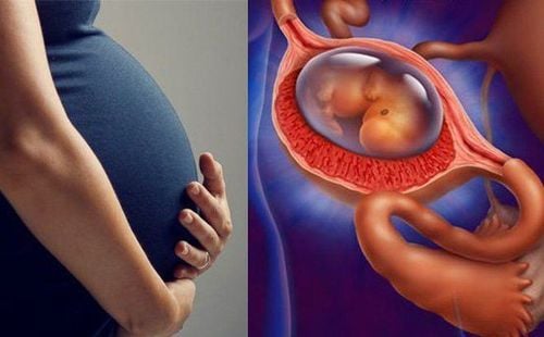
1.1. Month 1: Embryo develops three layers During embryonic and fetal development, most bones start out as flexible cartilage, which is gradually reshaped and transformed into a more rigid skeleton. Immediately after conception, the embryo differentiates into three layers of cells, namely:
The dermis: Also known as the middle layer, which will develop into the bones, heart muscle, kidneys and sex organs of the fetus. Endoderm: This is the inner layer that will become your baby's digestive system, liver, and lungs. Epidermis: This outer layer will develop into your baby's nervous system, hair, skin, and eyes. 1.2. February: Beginning of arms and legs Several major changes are taking place inside the tiny embryo, including:
Appearance of several types of bones Baby begins to develop basic parts of the collarbone and parts of the body backbone. At the same time, the neural tube - the foundation of the nervous system as well as the spine and skull, gradually formed.
Formation of arms and legs By about 6 weeks, the embryo also sprouts buds, which are the source of later arms and legs.
The tail disappears The only part that doesn't continue to grow is the tail similar to the tadpole of the embryo. Instead, they shrink and eventually disappear, leaving only the bones of the spine.
1.3. March: Face, nose, fingers and toes During the last weeks of the first trimester, fetal bone development takes place quite rapidly, including the fetal nasal bones.
The small buds now have the shape of arms and legs, along with flexible flexing joints. In addition, fingers and toes are well-defined around the 13th week. The upper extremities tend to grow strongly and are several days earlier than the lower extremities. This is similar to the skill development of a baby after birth, first moving the head, then rolling, turning, then crawling, and finally walking.
2. Second trimester
The fetus will absorb about 30 grams of this important mineral from the mother during 9 months to create more than 300 pieces of bones and joints of all kinds. If the required amount of calcium is not enough, calcium from the mother's bones is automatically secreted, sometimes leading to depletion or osteoporosis.
Therefore, mothers need to make sure to supplement with about 1,000 milligrams of calcium per day, not only for the bone development of the fetus but also for the protection of the pregnant woman herself.
2.2. May and 6: Baby moves limbs This is also the time when fetal bone development is active. At this stage, the fetus can wiggle its limbs and you will start to feel your baby's first movements around 18 weeks onwards.
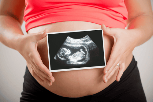
What does fetal femur length say? Through ultrasound images, the doctor will measure the length of the thigh bone connecting the baby's bottom to the knee. This index helps to assess and monitor whether the growth of the fetus is normal or not.
3. Third trimester
3.1. July and 8: Transforming cartilage into bone Entering the third trimester, most of the calcium that the mother adds from milk products will be provided to the baby. Thanks to about 250 milligrams of calcium per day, the fetus can focus on converting cartilage into bone, as well as building muscle and accumulating an extra layer of protective fat throughout the body.3.2. September: Fetal bones are still soft Week 36 is the peak period of the baby's absorption of calcium from the mother, with levels up to 350 milligrams/day from now until the end of pregnancy. However, the bones of a fetus are still softer than those of an adult. This feature helps the baby to move comfortably through the narrow birth canal at around 40 weeks.
In particular, the fetal skull is made up of many separate bone fragments, which can move flexibly and make the head flexible. baby's compression during labor. A newborn's bones will continue to grow stronger after the baby is born.
4. Infant bone development
As babies grow taller and stronger, all the bones in their bodies grow as well. After birth, the fetal bone growth no longer passes through the placenta, instead is the process of calcium absorption in the intestinal wall. For the skull bones, it usually takes 2-4 months for the anterior fontanelle to close and 18 months or more for the posterior fontanelle to close. This allows the baby's skull to expand and keep pace with the rapidly developing brain. Even after the fontanelles close, there will still be some empty space along the seam between the bones of the skull. Therefore, the brain and the entire head will not stop developing.When the child is about 2 - 3 years old, the joints of the bones begin to fuse together and the fetal skeletal development will continue until the growth plates fuse during puberty. At all stages of life, an adequate mineral intake is essential for bone growth and maintenance of strong properties with adequate strength.
5. Calcium supplements during pregnancy

Milk, soy milk; Dairy products: Cheese, yogurt; Canned salmon and tuna; Green vegetables, mushrooms and boiled soybeans; Egg; Cereals; Orange juice; You should consult a nutritionist if you want to take more calcium supplements. On the other hand, some data indicate that fetal bone development is also related to protein and parathyroid hormone (PTH).
To support the smooth development of fetal bones, mothers need to take vitamins regularly throughout pregnancy as well as continuously until breastfeeding. Newborns will get all the calcium they need from breast milk and formula, but if they're not exclusively breastfed, they need a vitamin D supplement. The calcium content must change to suit each stage, from infancy to weaning and even the long growth path in the coming future.
Vinmec International General Hospital offers a Package Maternity Care Program for pregnant women right from the first months of pregnancy with a full range of antenatal care visits, periodical 3D and 4D ultrasounds and routine tests to ensure that the mother is healthy and the fetus is developing comprehensively. In addition to regular check-ups, pregnant women will also be advised on nutrition and exercise so that the mother can gain weight reasonably while the fetus still absorbs nutrients well.
Doctor Nguyen Anh Tu has 6 years of experience in obstetrics and gynecology ultrasound, specially researched and trained in pregnancy ultrasound - prenatal diagnosis. Dr. Tu has completed courses on ultrasound - prenatal diagnosis of the FMF International Fetal Medicine Association; trained in consulting and implementing diagnostic intervention techniques in fetal medicine and participated in many specialized conferences and seminars on Fetal Medicine. Currently, he is a doctor at the Department of Obstetrics and Gynecology, Vinmec Hai Phong International General Hospital.
Please dial HOTLINE for more information or register for an appointment HERE. Download MyVinmec app to make appointments faster and to manage your bookings easily.
The article references the source: Whattoexpect.com;Ncbi.nlm.nih.gov; Embryology.med.unsw.edu.au


