This is an automatically translated article.
The article was written by MSc resident Nguyen Thanh Vinh - Obstetrician and Gynecologist, Department of Obstetrics and Gynecology - Vinmec Ha Long International General Hospital.Perineal tears at birth occur when the fetal head passes through the enlarged vaginal wall, in cases where the fetal head is too large for the stretch of the vaginal wall or the fetal head is normal but has poor elasticity of the vaginal wall. religion. The tear usually occurs in the skin and under the skin of the vagina and vulva, and usually heals on its own after a few weeks. In some cases the tear is deeper and requires treatment.
1. Classification of the degree of perineal tear
Depending on the damage to the layers of the vaginal area, people divide the tear into several levels:
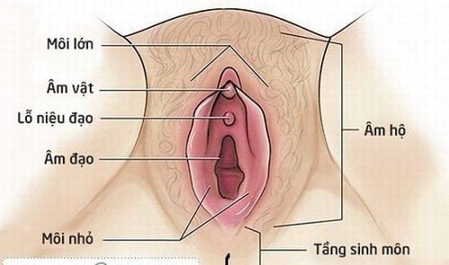
Grade 1 tear: the least serious, occurring only in the perineal skin - the skin around the vagina and between the vagina and the anus. You may feel slight pain or stinging while urinating. The tear does not require stitches and heals on its own after a few weeks. 2nd degree tears: are tears that include the skin and muscle of the perineum, which may extend up to the vaginal wall. These tears need stitches to heal in a few weeks. 3rd degree tear: tear into the perianal muscle – anal sphincter. This tear sometimes needs good pain relief in the operating room for the doctor to stitch to restore the anal sphincter. Recovery may need to be monitored longer than usual, as the most common postpartum complications in these cases are fecal incontinence and pain during sex that require monitoring. Grade 4 tear: is the heaviest level. The tear tears through the anal fascia into the rectal mucosa. This degree of tear requires a more specialized suture technique and is usually performed in the operating room. Incontinence and pain during sex are common complications and need to be monitored and detected early after birth.
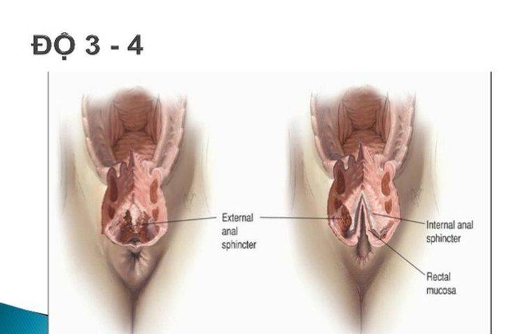
Rách tầng sinh môn cấp độ nặng
2. What is cervical tear?
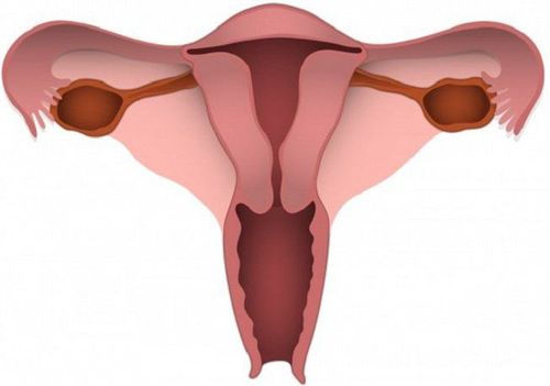
The uterus is an organ made up of thick layers of smooth muscle, which is the place where the embryo implants and nourishes and protects the fetus until birth. The uterus has a truncated cone shape, the base is wide at the top, the tip is turned down, divided into 3 parts: body, waist and cervix.
During labor, under the effect of uterine contractions, the cervix turns from a cylinder to a thin sheet - that is cervical effacement, which will be combined with cervical dilation during labor. uterus to form the lower uterine segment, facilitating the expulsion of the fetus. At this time, for some reason, women pushing to give birth too early or having surgical intervention due to difficult birth can lead to cervical tear complications.
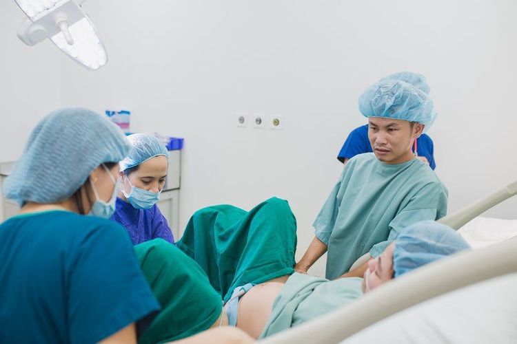
Rách cổ tử cung xảy ra trong khi sinh đẻ
Cervical tear can be a solitary lesion or be associated with perineal vaginal tear. In addition, cervical tear is classified according to the location of the tear in the circular plane passing through the cervix or the high and low position for the attachment to the vaginal canal.
If the cervical tear is located below the place where it is attached to the vaginal wall, the degree of injury is mild, and there is little bleeding. On the contrary, if the tear site is located on the attachment of the cervix to the vaginal wall, the damage is often severe, bleeding a lot, sometimes leading to shock, reducing the volume and affecting life.
Vinmec International General Hospital is one of the hospitals that not only ensures professional quality with a team of leading medical doctors, modern equipment and technology, but also stands out for its examination and consultation services. comprehensive and professional medical consultation and treatment; civilized, polite, safe and sterile medical examination and treatment space.
Customers can directly go to Vinmec Health system nationwide to visit or contact the hotline here for support.


