This is an automatically translated article.
Endotracheal anesthesia in renal drainage surgery is a method of general anesthesia, which helps control breathing and ensures patient safety.1. Endotracheal anesthesia in renal drainage surgery
Renal drainage is a common procedure, indicated to resolve acute purulent stasis in the kidney, caused by causes such as kidney stones, ureteral stones or acute renal failure due to bilateral ureteral stenosis...When performing renal drainage procedures, it is necessary to choose a safe method of anesthesia. Endotracheal anesthesia is the commonly chosen method of general anesthesia, helping to control breathing during and after kidney surgery for patients.
2. Indications and contraindications for renal drainage
Indicated in renal drainage due to causes such as:pyelonephritis due to kidney stones; Acute renal failure due to ureteral stricture. Contraindications:
Absolutely contraindicated when there is local infection of the drainage area. Relative contraindications when: The patient has a blood clotting disorder, does not agree, does not have enough anesthesia and resuscitation facilities or the operator is not technically proficient.
3. Procedure for performing endotracheal anesthesia in renal drainage surgery
3.1 Preparations Performers: Doctors, nurses specializing in anesthesiology and resuscitation.Facilities:
Anesthesia machine system with ventilator; hand squeeze oxygen source; monitor vital functions including electrocardiogram, arterial blood pressure, SpO2, EtCO2, temperature, breathing rate; defibrillator and phlegm aspirator. Laryngoscope, suction tube, endotracheal tube of different sizes, mask, squeeze balloon, oropharyngeal cannula, Magill pliers, soft mandrin. Medicines: Prepare drugs used in anesthesia and resuscitation and used to handle accidents, if any. Other means for prevention in difficult intubation cases such as: Cook tube, laryngeal mask, tracheostomy kit, flexible bronchoscope, mouth opener... Patient:
Anesthesiology examination before surgery to drain the kidney to detect and prevent possible risks during the procedure. Evaluation of difficult intubation. Use a sedative the night before surgery (if needed). Fasting 6 hours before surgery. Explain to family members and patients the risks of complications and what to watch out for to be able to cooperate during the procedure.
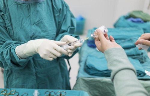
Người bệnh phẫu thuật dẫn lưu thận sẽ được gây mê toàn thân
Patient position: Lie on back, give 100% oxygen 3-6 l/min before starting anesthesia at least 5 minutes. Install a monitor to monitor signs such as electrocardiogram, SpO2, EtCO2... Create an effective intravenous line for the patient. Premedicate with medication if needed. Anesthesia:
Anesthesia can be initiated by intravenous anesthetics such as propofol, thiopental, etomidate... inhalation anesthetics (sevoflurane...) or a combination. Combine some drugs to increase the effect of anesthetics such as: opioid analgesics, muscle relaxants (rocuronium, vecuronium...) if necessary. Prophylaxis of gastroesophageal reflux with H2-receptor antagonists and metoclopramide. Conditions for intubation: Conditions for endotracheal intubation are when the patient is in deep sleep, with enough muscle relaxation in most cases. Oral or nasal intubation can be done.
Steps for oral intubation:
Open the patient's mouth, put one hand under the neck, insert the laryngoscope to the right side of the mouth, move the tongue to the left, push the lamp deep down, coordinate with the hand The rest press the cricoid cartilage, find the epiglottis and the glottis. Conduct rapid induction of anesthesia for the patient and perform the Sellick maneuver in cases of full stomach (press the cricoid cartilage with a force of about 20-30 kg as soon as the patient loses consciousness until the person performing the endotracheal intubation) accomplished). Pass the endotracheal tube gently through the glottis, stopping when the balloon of the endotracheal tube passes about 2 to 3 cm across the vocal cords. Gently withdraw the laryngoscope and inflate the endotracheal balloon. Check after removal of the laryngoscope for correct position of the endotracheal tube by listening to the lungs and EtCO2 readings. Fix the endotracheal tube with adhesive tape and place the cannula in the mouth to prevent the patient from biting the tube (if necessary). Nasotracheal intubation technique: Patients undergoing renal drainage surgery can intubate the nasal passages by following these steps:
Choose the side of the nose that is open and use vasoconstrictor drops (naphazoline, otrivin, etc.) . Choose a smaller endotracheal tube size than an oral endotracheal tube. Insert the lubricated endotracheal tube through the selected nostril. Open the patient's mouth, then proceed to insert the laryngoscope to the right side of the mouth, move the tongue to the left, push the lamp deeply, coordinate with the other hand to press the cricoid cartilage to find the epiglottis and glottis. Case if favorable: The performer gently intubates the endotracheal tube through the glottis and stops when the balloon of the endotracheal tube passes about 2 to 3 cm through the vocal cords. If it is difficult, use Magill pliers to clamp and guide the tip of the endotracheal tube to the correct position of the glottis; Need an assistant to push the endotracheal tube in from the outside. Gently withdraw the laryngoscope and then inflate the endotracheal balloon. Check after removal of the laryngoscope for correct position of the endotracheal tube by listening to the lungs and EtCO2 readings. Fix the endotracheal tube and place the cannula in the mouth to prevent the patient from biting the tube if necessary. In case the patient is difficult to intubate, it is necessary to apply the steps according to the difficult intubation procedure.

Có thể gây mê toàn thân bằng đặt nội khí quản đường miệng hoặc đường mũi
Monitoring the patient after general anesthesia:
Need to control the patient's breathing with a ventilator or hand squeeze. Monitoring the depth of anesthesia based on heart rate, arterial blood pressure, sweating, tears... Monitor vital signs by machine such as: Heart rate, blood pressure, SpO2, EtCO2, body temperature. Watch out for cases where the endotracheal tube is placed in the wrong position, or the endotracheal tube is folded or blocked. 3.3 Criteria for extubation When the surgery is completed, it is necessary to extubate the patient when the following criteria are met:
The patient is awake and follows orders; Self-elevating head for more than 5 seconds, TOF greater than 0.9 if any; The patient can breathe on his own regularly and the respiratory rate is within normal limits; Stable pulse and blood pressure; Body temperature over 35 degrees Celsius; There were no complications of anesthesia or surgery.
4. Complications and how to handle complications due to intubation
The process of endotracheal anesthesia in renal drainage surgery can occur some complications that need attention.4.1 Reflux of gastric juice into airway Signs: There is gastrointestinal fluid in the oral cavity and in the airway. Treatment: Remove the fluid immediately. Have the patient lie with the head low and to the side. Rapid endotracheal intubation and clear airway drainage; Monitor and prevent the risk of pneumonia after surgery. 4.2 Hemodynamic disorders Hypotension or hypertension, cardiac arrhythmias (bradycardia, tachycardia, arrhythmia). Depending on signs of hemodynamic disturbances to take measures. 4.3 Incident due to intubation Failure to intubate: Reintubation according to difficult intubation procedure or if not possible, need to switch to another method of anesthesia. Wrongly placed in the stomach Signs: Auscultation of the lungs does not see alveolar murmur, EtCO2 cannot be measured. Treatment: Reinsert the endotracheal tube. Vocal, air, and bronchial spasms Difficulty or non-ventilation presenting with auscultation with crackles or no alveolar murmurs. Management: Provide adequate oxygen, add sleeping pills and muscle relaxants, ensure ventilation and give bronchodilators, corticosteroids. If the patient's breathing cannot be controlled, a difficult intubation procedure should be applied immediately to ensure oxygen supply to the patient through the endotracheal tube. Trauma during intubation: Injuries can include bleeding, broken teeth, falling teeth into the airways, damage to the vocal cords, foreign objects falling into the airways... Diagnosis and treatment depend on each individual. Specific case. 4.4 Respiratory complications Folding or retracting the endotracheal tube, causing the tube to be pushed deep into one lung, opening the respiratory system or running out of oxygen...
Treatment: Ensure ventilation and provide adequate oxygen immediately. 100%, find and solve the cause.
4.5 Other complications of extubation Respiratory failure that occurs after extubation can occur for a variety of reasons. Need to provide oxygen to the patient immediately and find out the cause. Sore throat hoarse voice. Constriction of the vocal cords, trachea, and bronchi. Inflammation of the upper respiratory tract. Narrow larynx, trachea.

Đau họng là biến chứng có thể gặp sau khi rút ống nội khí quản gây mê toàn thân
In summary, surgical drainage of the kidney is often performed as an emergency treatment for patients with pus accumulation in the kidney causing renal failure. When conducting endotracheal anesthesia and surgery, many complications can occur, in order to limit the patient should perform treatment at reputable facilities. Currently, Vinmec International General Hospital is one of the addresses that strictly apply safe surgical anesthesia practice standards according to international guidelines. Vinmec has a team of experienced anesthesiologists and nurses, modern equipment such as: nerve detectors, ultrasound machines, Karl Storz difficult airway control system, comprehensive anesthesia monitoring system GE's AoA (Adequate of Anesthesia) including monitoring of anesthesia, pain and muscle relaxation will provide high quality and safety, helping patients to have adequate anesthesia, not wake up, and do not have residual muscle relaxants after surgery.
Vinmec Health System is also proud to be the first hospital in Vietnam to sign with the World Anesthesiology Association (WFSA) towards the goal of becoming the safest hospital for surgical anesthesia in Southeast Asia.
Please dial HOTLINE for more information or register for an appointment HERE. Download MyVinmec app to make appointments faster and to manage your bookings easily.