This is an automatically translated article.
The article is professionally consulted by Master, Doctor Nguyen Van Huong - Department of Diagnostic Imaging - Vinmec International General Hospital Da Nang.Magnetic resonance imaging (MRI) using magnetic contrast is the best imaging modality for evaluating meningiomas. Important advantages of MRI in imaging meningiomas are its superior resolution of various soft tissue types, its ability to render 3D, and its assessment of tumor contrast enhancement. This article will analyze the advantages of MRI in the diagnosis of meningiomas.
1. What is magnetic resonance imaging?
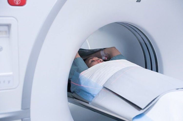
Magnetic Resonance Imaging (MRI) is a method of obtaining images of living organs by observing the amount of water inside each structure of the organ. The principle of magnetic resonance is based on a physical phenomenon called nuclear magnetic resonance.Besides, there is a difference compared to X-ray which uses radiation energy from X-rays, magnetic resonance imaging uses radio energy. Therefore, the risk of radiation contamination when taking magnetic resonance imaging is completely absent, while the image structure is reflected more clearly. Therefore, this diagnostic tool is preferred to be applied in the investigation of soft tissue diseases with complex or intricate structures.
2. The role of magnetic resonance imaging in the diagnosis of meningiomas

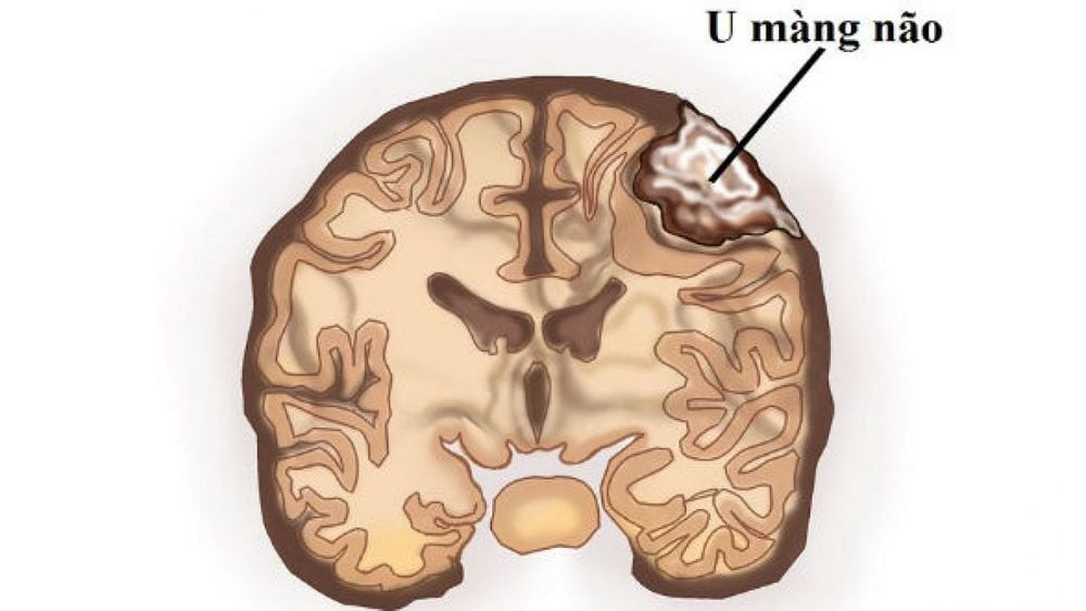
Hình ảnh u màng não
Magnetic resonance imaging can very clearly detect and characterize abnormalities in the brain parenchyma in general, as well as vascular tumors, arterial occlusions, venous sinus invasion as well as the relationship between tumor and surrounding structures.
Image of MRI in meningiomas, on T1-weighted images, most meningioma shows no difference in signal intensity compared with cortical gray matter. However, in some cases meningiomas may be noted to have a higher perfusion pressure than the cerebral cortex. In general, T1 imaging is mainly used to identify necrotic tumors, intratumoral hemorrhage, or tumors of a cystic nature.
Image in the T2W phase, the gain signal has changed completely, it is a fairly homogeneous gain block. Imaging is also helpful in evaluating hemorrhages and cysts. In addition, T2-weighted imaging helps to evaluate the CSF clearance between the neoplastic tissue (meningeal tumor tissue) and the brain parenchyma, confirming the tumor boundary. Moreover, the role of the T2W phase is to reflect the homogeneity of the soft tumor. This is seen more clearly in meningiomas, malignancies in general.
Signal intensity with T2 phase correlates very well with not only homogeneity but also tissue profile. In particular, with low-intensity signal, the tumor is fibrous and stiffer than normal parenchyma; for example, the tumor has a fibroblastic nature, while the more intense sections show a softer characteristic such as a vascular tumor.
For phase reversal recovery (FLAIR), this phase is very useful to assess the consequences of edema. Although this finding is not specific for meningiomas in particular, it is very meaningful in the diagnosis as well as the long-term prognosis for the patient.
Besides, similar to computed tomography, the "contrast" used in MRI is a gadolinium-based contrast agent; in which, meningiomas will be very evident in the injection phase. Several studies have reported that gadolinium injection helps detect meningiomas in more than 85% of cases. Furthermore, gadolinium also allows images of meningiomas to be more refined than those obtained without contrast. This is the basis for guiding the next surgical intervention process.

Nhìn chung, độ nhạy và độ đặc hiệu của MRI rất cao trong chẩn đoán u màng não
Overall, the sensitivity and specificity of MRI are very high in the diagnosis of meningiomas. MRI has been shown to be superior in delineating the tumor and its relationship to surrounding structures. Therefore, if there are any abnormal symptoms on the central nervous system such as headache, blurred vision, weakness..., it is necessary to consult a specialist and perform appropriate imaging techniques to be treated. Early treatment to prevent possible complications.
At Vinmec International General Hospital, magnetic resonance imaging (MRI) is used to diagnose meningiomas. Vinmec gathers a team of highly qualified and experienced neurologists; modern medical equipment, up to international standards; professional service quality, helping to improve the efficiency of disease diagnosis and treatment.
Master Doctor Nguyen Van Huong used to be a Lecturer in Medical Imaging at Danang University of Medicine and Pharmacy, many years of experience working in hospitals such as Cancer Hospital; Da Nang Hospital; Da Nang Obstetrics and Gynecology Hospital.
For detailed advice on meningiomas and diagnostic methods, please come directly to Vinmec health system or register online HERE.
References: medscape.com
MORE:
Structure of the meninges What are the benefits of Magnetic Resonance Imaging (MRI)? What is the effect of magnetic resonance imaging (MRI) of the brain? Brain tumors in children: Causes, symptoms, diagnosis and treatment


