This is an automatically translated article.
The article was professionally consulted with Specialist Doctor II Lai Thi Nguyet Hang - Obstetrician and Gynecologist - Department of Obstetrics and Gynecology - Vinmec Ha Long International General Hospital.Benign fibroadenoma, although not dangerous to the patient, will create discomfort and affect aesthetics, the signs of the disease are often difficult to distinguish from breast cancer.
1. How is breast cancer diagnosed?
About 50% of women will develop fibroadenoma at some point in their lives. Bilateral breast clinical examination can detect a mass if it is relatively large in size. Some fibroids are too small to feel, so they can only be detected on imaging tests.If a palpable lump is found, your doctor may recommend certain tests that are appropriate depending on your age and the characteristics of the tumor. Tests to evaluate for fibrocystic breast tumors include:
A mammogram to create pictures (mammograms) of suspicious areas in the breast tissue. A fibroma may show up on a mammogram as a breast mass with smooth, rounded edges that is distinct from surrounding breast tissue. Breast ultrasound: This technology uses sound waves to create an image of the inside of the breast. Your doctor may recommend a breast ultrasound in addition to a mammogram to evaluate for a breast lump if you have dense breast tissue. If a mammogram shows a lump in the breast or other abnormality, a breast ultrasound may be used to further evaluate the tumor. A breast ultrasound can help your doctor determine if a fibrocystic breast lump is solid or filled with fluid. A solid mass is more likely to be a fibroadenoma; a fluid-filled mass is more likely to be a cyst. Fine-needle aspiration: A thin needle is inserted into the tumor to assess tumor characteristics. U.S. biopsy. A radiologist with guidance from an ultrasound usually performs this procedure. The doctor uses a needle to collect tissue samples from the tumor, which is sent to a laboratory for histopathological analysis of the tumor. Helps differentiate from breast cancer.

2. How is breast cancer treated?
Fibroids in many cases do not require treatment. However, some women choose to have a mastectomy for peace of mind. If it is certain that the breast lump is fibrocystic based on the results of clinical breast exams, imaging tests and biopsies, surgery may not be necessary because:Surgery can distort shape and texture of the breast Fibroids sometimes shrink or disappear on their own. Breasts with many fibroids appear stable and do not change in size on ultrasound compared with previous ultrasound If no surgery is chosen, it is important to monitor the tumor fibrocystic breast disease with follow-up visits to your doctor for breast ultrasounds to detect changes in the appearance or size of a tumor.
3. Surgery to remove fibrocystic breast in which case?
3.1 Indications and contraindications The doctor may recommend fibrocystectomy in the following cases:Breast fibroids are too large, larger or cause symptoms The fibrocysts are benign or the risk of deterioration leads to bad results. to cancer Patients who wish to have fibrocystectomy are contraindicated in the following cases:
Suspicious lesions are more unusual Patient has not had children After removal of fibroids, there may be a or multiple new fibroadenomas develop. New breast lumps should be evaluated with a mammogram, ultrasound, and possibly a biopsy to determine if the lump is a fibroid or possibly cancerous.

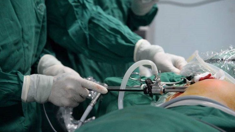
Step 2: Determine the location of the fibrocystic breast tumor to be removed, if it is small, a needle can be used to determine the best landmark. Under anesthesia, if not available, local anesthesia. After making an incision through the skin and subcutaneous tissues, use scissors to dissect to go straight into the tumor to avoid crushing surrounding tissues and causing bleeding. Remove the tumor through the incision after dissecting and carefully stopping the bleeding of the tissues around the tumor with the indicator. If the tumor is deep, then it is necessary to sew up the incision tissue after carefully checking and not see any bleeding.
Step 3: Stitch to restore the skin with sutures or suture under the skin with Vicryl 2.0 criteria.
Step 4: Cover the incision, which can be covered with an elastic bandage around the chest, if it is suspected that the dissection may still bleed, it will be removed after 12-24 hours. After the removal is complete, the removed tissue must be sent for histopathological examination. Using extra pain relievers and antibiotics and anti-edema drugs for patients
Vinmec International General Hospital offers a breast cancer screening service package that helps customers check and screen for signs of breast cysts as well as the risk of breast cancer. In the examination package will include methods of mammography and bilateral breast ultrasound diagnosis for accurate results, assisting doctors in the examination.
Please dial HOTLINE for more information or register for an appointment HERE. Download MyVinmec app to make appointments faster and to manage your bookings easily.


