This is an automatically translated article.
The article is professionally consulted by Master, Doctor Ly Thi Thanh Nha - Department of Obstetrics and Gynecology - Vinmec International General Hospital Da Nang. Doctor Nha has strengths and experience in fetal malformation ultrasound, 3D, 4D fetal ultrasound.Fetal echocardiography is an imaging technique that helps detect heart defects, if any. All pregnant women should have this test done during pregnancy as ordered by their obstetrician. So at which week of pregnancy is an echocardiogram most appropriate?
1. Beware of congenital heart defects
According to the World Health Organization (WHO), among children born, about 0.8 - 1% may have congenital heart disease. In which, about 25% of severe cases require surgical intervention.Congenital heart disease (CHD) is a common neonatal disorder with a high mortality rate. During the first 3 months, heart defects are very likely to cause death. If there is no death, the child's life will also have many accompanying disorders such as mental retardation, bronchitis or recurrent pneumonia, cardiac arrhythmias...
Treatment of existing congenital heart diseases Now there are many different methods such as medication, surgery or heart transplant. Some cases cannot be completely treated and greatly affect the child's later life.
In Vietnam, statistics from the Ministry of Health show that there are about 8,000 - 10,000 babies with various congenital heart diseases. However, only about half have surgery, the rest have to live with the disease for the rest of their lives. The reason for these sad numbers is that pregnant women subjectively do not have regular antenatal check-ups, especially not performing fetal echocardiography.
Therefore, pregnant women should pay attention to periodic antenatal check-ups to detect dangerous heart defects early through supportive paraclinical tests, the most important being fetal echocardiography.

2. What is fetal echocardiography?
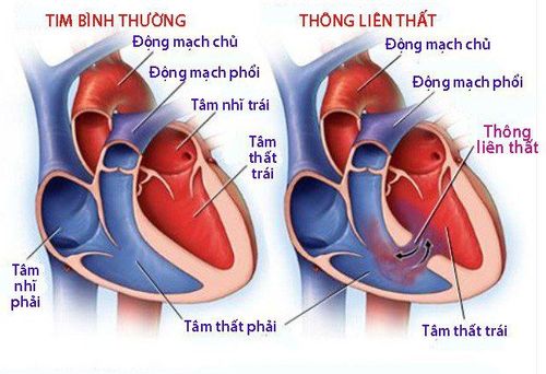
In Vietnam, many medical facilities have deployed fetal echocardiography to diagnose heart defects during pregnancy. However, the importance and significance of fetal echocardiography is still a big question mark for pregnant women, leading to subjective psychology and ignoring this important imaging test.Fetal echocardiography is a diagnostic imaging tool that helps evaluate fetal heart conditions such as: heart structure, characteristics of heart valves, shunt holes in the heart, heart rate...
With the ability to detect With congenital heart defects of the fetus right from the period in the womb, fetal echocardiography is recommended by doctors early to diagnose promptly and have appropriate treatment.
Today, modern ultrasound equipment combined with the high level of expertise of technicians has improved the accuracy of fetal echocardiography, which can detect congenital heart defects with high accuracy. Accuracy up to 99%.
3. Fetal echocardiography at which week?
Which week fetal echocardiography gives the most accurate results is the question of many pregnant women when being consulted to do this test.In most cases, fetal echocardiography to diagnose congenital heart defects should usually be performed at 17 and 18 weeks (or later to 22 weeks) for the most accuracy.

Because the baby is still quite small, a high-end 4D ultrasound system like the GE Voluson E10 will play a very important role in helping doctors at Vinmec detect more than 95% of anomalies during this period. Packed with the latest technologies, the GE Voluson E10 enables enhanced image quality and penetration for outstanding high-resolution images and easy operation.
4. Benefits of fetal echocardiography during pregnancy
Fetal echocardiography by different methods such as 2D - 3D - 4D ultrasound or color Doppler ultrasound... all help to assess the fetal heart condition, detect congenital anomalies, if any. Thereby, fetal echocardiography significantly reduces morbidity and mortality from congenital heart disease in children. In addition, the ultrasound detects abnormalities early to help doctors give appropriate treatment depending on the status of the fetal heart.In addition, fetal echocardiography is also a method of psychological stabilization of couples, helping them to prepare economically as well as measures for care and treatment when the baby is born.
5. Which pregnant women need to perform fetal echocardiography?
Fetal echocardiography is an important test during pregnancy. Some cases of pregnant women need periodic fetal echocardiography to monitor fetal development, including:Pregnancy after artificial insemination. There is a family history of congenital heart disease. Pregnant women with diseases such as phenyl ketonesuria, diabetes, ... or other genetic diseases such as: Marfan, Ellis Van Creveld, Noonan... Pregnant women infected with Rubella virus.

Fetal heart arrhythmia; Multiple pregnancy or twin-twin transfusion syndrome; Abnormal nuchal translucency; Other accompanying abnormalities such as umbilical hernia, diaphragmatic hernia, duodenal atresia... Fetal echocardiography is a routine test for doctors to assess fetal heart status, heart rate and detect abnormalities. usually fetal heart. However, this diagnostic technique requires the operator to have good expertise, in-depth experience and well-trained, because there are many cases of heart defects that have been missed, losing opportunities. detection and treatment. In addition, pregnant women should also choose reputable antenatal clinics that have standard and modern ultrasound systems to have clear and realistic images.
Fetal echocardiography is a routine test performed at Vinmec International General Hospital. Accordingly, at Vinmec, many cases of pregnant women with fetuses with very severe heart defects have been diagnosed and detected, and at the same time, surgical intervention has been performed to treat them early from the day they are born, providing a high prognosis for survival. for many babies, bringing joy and happiness to many families.
Therefore, to ensure the health of both mother and fetus, right from the start of pregnancy, mothers can refer to the maternity package package at Vinmec International General Hospital. There is a team of well-trained and specialized medical doctors, capable of detecting birth defects of the fetus very early. Accordingly, depending on the stage of pregnancy, the pregnant woman will be consulted by the doctor for an appropriate pregnancy ultrasound to check the development status of her baby.
For more information, please contact the hospitals and clinics of Vinmec Health system nationwide.
Please dial HOTLINE for more information or register for an appointment HERE. Download MyVinmec app to make appointments faster and to manage your bookings easily.


