This is an automatically translated article.
Articles by Master, Doctor Phan Van Phong - Emergency Resuscitation Department - Vinmec Central Park International General Hospital
PiCCO is a hemodynamic exploration technique based on the principle of transpulmonary thermodilution, which is highly applicable, fast, with few complications and can be applied in emergency departments.
1. Learn about PiCCO
PiCCO (pulse contour cardiac output) is an advanced hemodynamic monitoring system. The operating principle is based on the combination of the transpulmonary thermodilution method and the continuous pulse contour analysis method, which measures continuously and simultaneously many hemodynamic parameters such as CO, pre-amplification. burden, systemic resistance, cardiac contractility, and extravascular volume in the lungs without the need for right-heart catheterization.
2. Indications and contraindications when performing PiCCO . technique
2.1. Indications Patients with hemodynamic instability: Shock, Acute Heart Failure, Anaphylaxis, Acute Pulmonary Injury (ALI), Adult Advanced Respiratory Failure (ARDS) Multiple Trauma, Severe Burns, Multiple Failure organs. Patients at high risk in major interventions such as organ transplantation, cardiovascular surgery, major abdominal surgery... 2.2. Contraindications This method is contraindicated for arterial and venous catheterization such as: not indicated for femoral artery bypass surgery in the groin area (femoral artery transplant) or severe bilateral inguinal burns. Alternative arterial routes (axillary, brachial, radial arteries) may be used.
3. Some limitations of the PiCCO . method
PiCCO technique may give inaccurate results in the following cases: Patient has a large flow between the right and left hearts Running extracorporeal circulation Aortic aneurysms Severe arrhythmias Cases where a large part of the lung is removed , or pulmonary infarction If the arterial curve is poor quality, the value of CO measured by pulse wave method will be inaccurate.
4. Clinical application of PiCCO . technique
4.1. Cardiac output Cardiac output is an important parameter to evaluate hemodynamic disorders and guide treatment. All studies have shown that PiCCO pulse oximetry continuous CO measurement is reliable and accurate even in hemodynamically unstable conditions and is not affected by the use of drugs that alter blood pressure and strength. system circuit breaker.
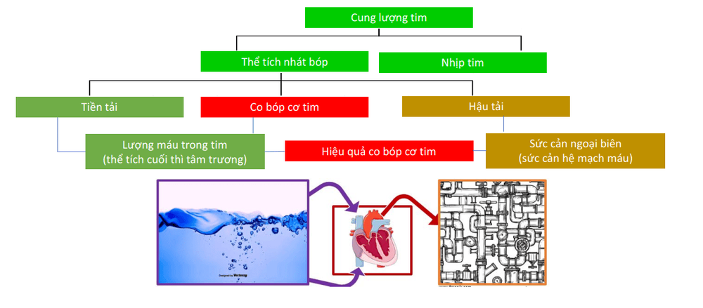
4.2. Preload Many studies have confirmed that ITBV and GEDV have a higher accuracy than chamber filling pressure in assessing preload.
Cardiac filling pressures (Central Venous Pressure: CVP and Pulmonary Obstructive Pressure - PAOP) are commonly used to assess preload, but those parameters are imprecise. Because end-diastolic ventricular pressure is dependent on chamber relaxation, it does not faithfully reflect preload volume. On the other hand, CVP and PAOP are affected when the patient is on mechanical ventilation, while ITBV and GEDV are not affected much by artificial ventilation
Some other indicators can be used to assess preload such as: terminal right ventricular volume diastole measured by pulmonary artery catheter; left ventricular end-diastolic area measured by echocardiography; ITBV measured by double indicator dilution method; and GEDV measured by the transpulmonary heat dilution method. However, the transpulmonary heat dilution method has more advantages, without the need for a pulmonary artery catheter, compared with measuring left ventricular end-diastolic area by echocardiography, GEDV does not depend on the level of the sonographer. can be carried out frequently and easily at the hospital bed.
4.3. Evaluating the response to fluids One of the most common challenges facing the resuscitator is the response of CO to an increase in circulating volume, which is a fact that most studies only show. found that only 50% of patients in severe condition respond to fluids, and in a few others, infusions are harmful to the lungs and other organs. Because the correlation between preload and stroke volume depends on ventricular contractility. Isolated assessment of ventricular preload is not sufficient to predict response to perfusion.
Although increasing or decreasing variation of preload volume parameters is used in predicting fluid response, in cases where the measured values are within the normal range, they will not what to predict. Therefore, a variety of hemodynamic parameters have been applied to evaluate the effect of fluid on hemodynamics, especially in mechanically ventilated patients with the effect of positive airway pressure on stroke volume. In patients with sedation, positive pressure ventilation during inspiration changes SVV and PPV. Similarly, since vascular pressure is proportional to left ventricular stroke volume, and the variability of pulse pressure due to ventilation is closely related to stroke volume, it is possible to predict cardiac response to perfusion.
PiCCO system automatically calculates PPV and SVV in each stroke by analyzing pulse waves. Based on PPV and SVV, it is possible to predict the response of CO to fluid infusion in heart surgery patients. However, it is important to keep in mind that PPV and SVV are affected by tidal volume, e.g. during mechanical ventilation with high tidal volumes (>15ml/kg) the measured tidal volumes are lower than they actually are. economic.
Monitoring fluid adjustment according to SVV and PPV is a good method to assess the fluid response in patients using sedation and mechanical ventilation, suitable for patients under anesthesia during surgery, this is a great step forward in monitoring. track BN.
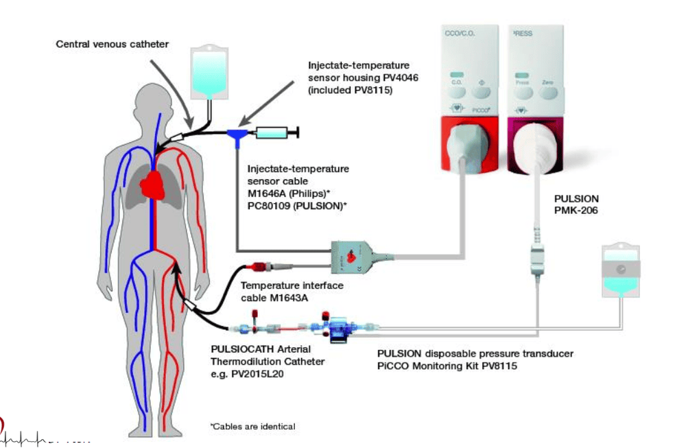
4.5. Evaluation of myocardial contractility In low blood flow conditions, myocardial contractility measurement may be useful to identify whether the patient is responding to inotropes. Accurate assessment of myocardial contractility at the bedside is not simple, as it depends on many factors, including preload and/or afterload. The ventricular ejection fraction is commonly used to assess the contractile function of the heart, which is the ratio of stroke volume to the left ventricular end-diastolic volume. Pulmonary thermal dilution measures GEDV, so the quotient between stroke volume and one-quarter of GEDV can estimate the total ejection fraction of the heart.
These parameters are calculated by the PiCCO method and can be used to assess whether the patient has impaired ventricular function or not. In addition, PiCCO can continuously assess left ventricular contractility by measuring dP/dtmax during the indicator injection phase.
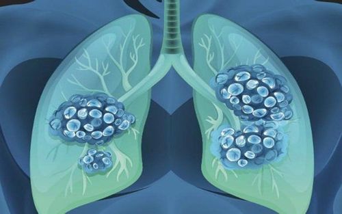
4.6. Determination of pulmonary edema and pulmonary vascular permeability Although chest X-ray and blood gas testing play a major role in the diagnosis of ALI and ARDS, these parameters have proven to be of little value in the diagnosis of pulmonary edema. diagnose pulmonary edema. Therefore, many methods are applied to evaluate pulmonary edema such as computed tomography, magnetic resonance imaging, chest wall resistance measurement, and heat dilution method.
The heat dilution method is a simple and highly sensitive method of measuring EVLW that can recognize 10-20% variation in EVLW, which occurs in the early stages of pulmonary edema when where clinical symptoms and other diagnostic signs have not yet appeared.
The EVLW parameter is of great value in guiding fluid administration especially in patients with increased permeability of small pulmonary vessels (eg, infection). The PiCCO system measures EVLW along with CO, preload (GEDV) and fluid response (PPV and SVV) parameters that can guide fluid therapy and especially in difficult-to-assess situations. prices, helping to reduce mortality.
Please dial HOTLINE for more information or register for an appointment HERE. Download MyVinmec app to make appointments faster and to manage your bookings easily.


